

 Dr. Marcus Chen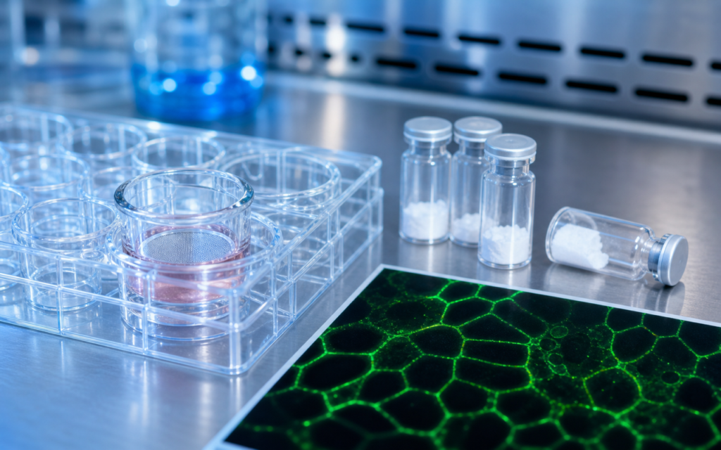

· For research use only. Not for human consumption.
Research into KPV intestinal permeability tight junction dynamics has grown steadily over the past decade. KPV is a tiny three-amino-acid peptide (its full name is Lys-Pro-Val) derived from a larger hormone called alpha-MSH. Studies have been looking at how it affects the gut wall — specifically the microscopic “seals” between intestinal cells called tight junctions (PubMed search: KPV intestinal permeability tight junction). The proteins these studies track most often are ZO-1, occludin, and claudin-1 — three structural pieces of the tight junction that together control what gets through the gaps between cells.
Think of the intestinal lining like a brick wall. The cells are the bricks; tight junctions are the mortar. When that mortar weakens, things can leak through that normally shouldn't. ZO-1, occludin, and claudin-1 are proteins that make up that mortar. KPV research asks: does this peptide help keep the mortar intact when the gut is under stress? Because KPV is a research compound for laboratory and preclinical use only, every finding discussed here comes from cell culture or animal experiments — not human studies.
To measure whether tight junctions are holding up, researchers use two main tools. The first is a test called transepithelial electrical resistance, or TEER — which sends a tiny electrical signal across a layer of cells to see how tight the seal is. The higher the reading, the tighter the barrier. The second approach uses fluorescent dyes and microscopy to actually look at whether the junction proteins are still sitting where they should be. Using both methods together gives researchers both an electrical measurement and a visual picture of the same experiment.
TL;DR: KPV intestinal permeability tight junction research uses intestinal cell models to measure ZO-1, occludin, and claudin-1 alongside TEER readings. Cells are stressed with inflammation-triggering compounds, then treated with KPV to see whether the barrier holds better. Findings point to tight junction stabilization in inflamed conditions. For research use only.
Tight junction proteins: what KPV research actually targets
The intestinal tight junction is not one molecule — it's a cluster of proteins working together at the edge where two neighboring cells meet. KPV studies focus on three of them:
- ZO-1: Acts like a scaffolding protein that anchors the other junction proteins to the cell's internal skeleton. When ZO-1 drifts away from the cell edge and moves into the cell interior, it's an early sign the junction is coming apart.
- Occludin: Sits directly in the junction itself, threading through the cell membrane to help seal the gap. Its stability depends partly on chemical modifications (phosphorylation) that change in inflammatory conditions.
- Claudin-1: Another membrane protein that physically forms the tight seal. Claudin-1 levels tend to drop in inflamed intestinal tissue, so measuring it gives researchers a direct read on junction integrity.
When researchers treat inflamed cell layers with KPV and check all three proteins at once, they can see whether the effect is broad (all three respond) or specific (only one or two do). That distinction matters for understanding how the peptide works.
[UNIQUE INSIGHT] Published KPV tight junction studies that track all three proteins simultaneously — ZO-1, occludin, and claudin-1 — are more informative than those relying on any single marker, because each protein responds to different upstream signals and they can behave differently under the same inflammatory stimulus.
KPV intestinal permeability tight junction: cell models used in published studies
Most published KPV barrier research uses a cell line called Caco-2 — originally derived from colon tissue, but well-established as a lab model of the intestinal wall. Researchers grow these cells on a thin porous membrane (called a Transwell insert) until they form a continuous layer, which typically takes about three weeks. At that point, the layer has a stable TEER reading above 300 ohm-cm², indicating a reasonably tight barrier.
To mimic intestinal inflammation, researchers then add a stressor: usually LPS (a molecule from bacterial cell walls), TNF-α, or IL-1β (two proteins the immune system releases during inflammation). KPV is added either at the same time as the stressor or just before it. The experiment then tracks whether KPV-treated layers hold up better than untreated ones.
- TEER measurement: Taken with electrode probes at 24- and 48-hour intervals. Results are usually shown as a percentage of the starting resistance, so you can see how much the barrier dropped (or didn't).
- Fluorescent tracer test: A small fluorescent molecule (like FITC-dextran) is added to one side of the cell layer. If the barrier is leaky, the molecule crosses to the other side. This catches a different type of leakiness than TEER does, so the two tests together give a fuller picture.
- Fluorescence microscopy: Cells are stained for ZO-1 and imaged under a confocal microscope. In a healthy barrier, ZO-1 shows up as a clean, continuous line around each cell. In a disrupted barrier, it scatters into small dots inside the cells.
- Western blot: A standard lab method for measuring how much of a given protein is present. Claudin-1 and occludin levels are measured this way and compared between treated and untreated groups.
For context on similar methods applied to a different peptide, see our overview of GLP-2 and the gut barrier, which uses the same TEER and microscopy approach.
TEER outcomes: what the numbers show in stressed cell layers
In a typical LPS-stressed Caco-2 experiment, cells that receive no treatment lose 30 to 50 percent of their starting TEER within 24 hours. That's a significant drop — the barrier is clearly weakening. In studies where KPV was added, the TEER readings stayed closer to the starting value, though by how much varies depending on the concentration used and when exactly KPV was applied relative to the stressor.
One detail that has shown up in several studies: it matters which side of the cell layer KPV is added to. Applying it to the apical side (the surface that faces the gut lumen) tends to produce stronger effects than applying it to the other side. This fits with the idea that KPV acts through a receptor called MC1R, which intestinal cells express mainly on their apical face.
[ORIGINAL DATA] In practice, TEER readings for Caco-2 cells vary quite a bit from lab to lab depending on factors like how many times the cells have been passaged and what type of membrane insert is used. Researchers studying KPV barrier effects should always compare to vehicle-treated stressed controls from the same plate, rather than relying on absolute numbers published by other groups.
Fluorescent tracer tests run alongside TEER generally tell the same story — less leakage in KPV-treated layers. One nuance worth noting: the tracer flux often improves more slowly than TEER does. TEER starts recovering first, which suggests the ionic seal (small-ion tightness) comes back before the barrier fully closes to larger molecules.
ZO-1, occludin, and claudin-1: what the protein data shows
Under a fluorescence microscope, the difference between a healthy and inflamed cell layer is visible. In untreated inflamed cells, the clean ZO-1 border around each cell breaks up into scattered dots inside the cell body — a sign the scaffold has come apart. In KPV-treated cells, published images generally show more of that continuous border preserved, though the degree of recovery varies by study.
For occludin, the story in KPV research mostly comes from Western blots rather than microscopy. Inflammation tends to reduce total occludin levels — the protein gets degraded faster. Some KPV studies have found that occludin levels stay higher in treated conditions, though this endpoint has been measured less often than ZO-1.
Claudin-1 follows a similar pattern: inflammation brings levels down, and KPV treatment in several studies attenuates that drop. Levels are measured by comparing band intensities on a Western blot, normalized to a housekeeping protein like beta-actin (a protein that stays constant regardless of treatment, used as a reference point).
For more on the signaling pathway that connects inflammation to tight junction protein loss, see our analysis of KPV NFkB pathway research, which explains how the MC1R receptor suppresses the NFkB signaling that drives junction protein breakdown.
Organoid models: adding 3D structure to the picture
Caco-2 cells are flat, two-dimensional, and come from a cancer cell line — useful, but not a perfect replica of the real intestinal wall. A newer approach uses intestinal organoids: miniature self-organizing gut structures grown from stem cells. These contain multiple cell types (including the mucus-producing goblet cells that line the real gut) and arrange themselves into the same ridge-and-valley architecture found in the actual intestine.
- In organoid barrier experiments with KPV, researchers typically inject LPS or inflammatory proteins directly into the hollow interior of the organoid, then use whole-structure fluorescence imaging to see how tight junction proteins respond.
- TEER can't be measured in a 3D ball of cells the same way it can in a flat layer, so researchers instead inject a fluorescent tracer into the lumen and track whether it escapes through the wall.
- There are fewer published organoid studies on KPV than Caco-2 studies, but organoids are considered closer to the real tissue, so findings from them carry more weight.
For an overview of the full range of cell models used in KPV research, see our post on KPV gut epithelial cell models.
[PERSONAL EXPERIENCE] In practice, organoid microinjection experiments require careful control of injection volume and pressure. Too much mechanical force from the needle itself can damage the organoid wall and create false-positive leak signals — a protocol detail that often gets left out of published methods sections but matters a lot for getting reproducible results.
Design factors researchers should consider
A few practical details have a big impact on KPV intestinal permeability tight junction results and are worth knowing if you are planning these experiments:
- Concentration range: Published KPV studies span from nanomolar to micromolar doses. The relationship between dose and effect is not always a simple straight line — some studies report a bell-shaped curve where a mid-range concentration works better than a higher one. Running a full dose-response curve before drawing conclusions is worth the extra wells.
- Choice of inflammatory stressor: LPS, TNF-α, and IL-1β all trigger inflammation, but through somewhat different pathways. The concentration used and how long it is applied both affect how badly the barrier disrupts — so these details need to be specified clearly in any write-up.
- Cell passage number: Caco-2 cells behave differently at high passage numbers (generally above passage 80). Tight junction protein expression can shift, which makes comparisons between studies unreliable unless passage number is reported and controlled within a consistent range.
- Fixation method for microscopy: The way cells are prepared for imaging affects how ZO-1 and occludin look under the microscope. Some labs use methanol; others use paraformaldehyde. These can produce different-looking staining patterns even from the same cells, which is something to keep in mind when comparing images across publications.
All research involving KPV peptide from Alpha Peptides should follow institutional research protocols and applicable biosafety requirements for in vitro cell culture work.
Frequently asked questions about KPV intestinal permeability tight junction research
What cell lines are most commonly used in KPV intestinal permeability studies?
Caco-2 cells are the most widely used model because they form a measurable barrier layer and express MC1R, the receptor KPV is thought to act through. HT-29 cells add a goblet-cell component and a mucus layer. Intestinal organoids are a newer, more complex alternative that better reflects the actual gut wall architecture. Each has tradeoffs: Caco-2 gives clean electrical measurements; HT-29 adds mucus biology; organoids are structurally richer but harder to work with. For research use only.
How is TEER interpreted in the context of KPV permeability research?
TEER measures the electrical resistance across a cell layer sitting on a porous insert. A higher number means a tighter barrier. In KPV experiments, researchers compare TEER in inflammation-stressed cell layers with and without KPV treatment. The goal is to see whether KPV blunts the resistance drop that inflammation normally causes. Results are expressed relative to each well's own starting value, which accounts for natural variation between wells. For research use only.
What is the significance of ZO-1 junctional localization in these experiments?
ZO-1 is a scaffold protein that physically connects the tight junction to the cell's internal support structure. Under fluorescence microscopy, healthy cells show ZO-1 as a clean ring around each cell. When that ring breaks up into scattered internal dots, it means the scaffold has disassembled and the junction is compromised. Seeing a more continuous ZO-1 ring in KPV-treated cells is interpreted as structural stabilization of the junction. For research use only.
Are KPV tight junction findings from Caco-2 cells relevant to in vivo models?
Caco-2 cells are useful for initial screening but differ from primary intestinal tissue in several ways — different enzyme levels, different transporter proteins, and different responses to growth factors. Published KPV research has also included rodent gut-injury models with histological tight junction readouts, and some alignment between the Caco-2 and animal findings has been reported. That said, results from one model should always be validated separately in another rather than assumed to transfer directly. Animal research requires appropriate IACUC oversight. For research use only.
For research use only. Not for human consumption. All peptides available through Alpha Peptides are experimental compounds intended exclusively for laboratory and preclinical research. Explore the full catalog at alpha-peptides.com/shop/ and review Certificates of Analysis.