

 Dr. James Kowalski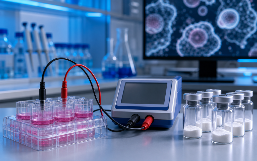

· For research use only. Not for human consumption.
KPV gut epithelial cell model research is one of the cleaner, more reproducible ways researchers study what this small peptide actually does inside the gut lining. KPV is a three-amino-acid chain (Lys-Pro-Val) that shows up in published preclinical work targeting receptors found along the intestinal wall (PubMed search: KPV peptide intestinal epithelial). Labs studying it have settled on three main cell systems: Caco-2, HT-29, and mini-gut structures called organoids. Each one answers a slightly different question about how the gut lining behaves, and picking the wrong one for a given experiment means the data won’t hold up. For background on how KPV signals through its receptor targets, see how KPV works and the introductory profile at what is KPV.
Part of why KPV works well in these lab setups comes down to its size. Three amino acids is tiny. A bigger molecule has a harder time reaching receptors on the cell surface without getting tangled up or sticking to the wrong things. KPV is small enough to reach those surface receptors quickly, which makes it easier to generate clean, repeatable dose-response data. This article walks through each cell model used in published KPV gut research, what the experiments actually measure, and the practical mistakes that tend to make results fall apart between labs. All discussion covers preclinical in vitro research only. For research use only. Not for human consumption.
TL;DR: KPV gut epithelial cell model research uses two types of flat cell layers (Caco-2 and HT-29) alongside three-dimensional mini-gut structures (organoids) to track how this small peptide affects the gut lining’s tightness and its output of inflammatory signals. The main measurements are electrical resistance across the cell layer and levels of inflammatory proteins released into the surrounding fluid. For research use only.
Caco-2 cells: the standard barrier model in KPV gut epithelial cell model research
Think of the gut lining as a wall of bricks held together by mortar. The “bricks” are epithelial cells. The “mortar” consists of proteins called tight junctions that seal the gaps and stop unwanted material from leaking through. Caco-2 cells are a lab-grown human cell line that naturally forms this same brick-and-mortar structure when given about three weeks to mature on a porous membrane. That makes them the go-to starting point for KPV gut epithelial cell model research focused on whether the gut wall stays tight or starts to leak.
The main measurement researchers use here is called transepithelial electrical resistance, or TEER. It works like this: a weak electrical current passes across the cell layer, and the resistance it meets tells researchers how intact the barrier is. A tight, healthy layer resists more current. A leaky one resists less. When researchers chemically inflame the Caco-2 layer (using bacterial byproducts or inflammatory proteins like IL-1β and TNF-α), TEER typically drops 30–60% within 24–48 hours. Researchers then apply KPV to see whether that resistance drop is slowed, prevented, or partially reversed.
One detail that matters a lot: when exactly KPV gets added. Adding it before the inflammation, at the same time, or after produces different results. Those aren’t interchangeable experiments — each one models a different real-world scenario. Labs that don’t record this timing precisely end up with data that can’t be compared to anyone else’s.
[UNIQUE INSIGHT] Caco-2 cells carry low but detectable levels of the MC1R receptor (the surface protein KPV binds) after they fully mature. But the amount varies with how many times the cells have been passaged (split and re-grown). Researchers should confirm MC1R levels by gene expression or protein analysis in the exact cell batch they’re using before concluding that TEER changes are driven by receptor engagement specifically.
HT-29 cells: a mucus-producing complement to Caco-2
Caco-2 cells are good at simulating the gut wall’s electrical barrier, but they produce almost no mucus. The real gut lining is coated in a layer of mucus that acts as a first line of defense. HT-29 cells (and a closely related variant called HT-29-MTX) do produce mucus, which adds a more realistic dimension to KPV gut epithelial cell model research when the question involves how KPV navigates that mucus coating to reach underlying receptors.
HT-29 cells aren’t great for the resistance measurement (their barrier is leakier by nature), so researchers use them primarily to measure inflammatory protein output. The typical setup: inflame the cell layer with IL-1β or TNF-α, then measure the inflammatory proteins those cells release into the surrounding fluid. The main proteins tracked are:
- IL-8 (CXCL8): the most reliable signal; inflamed HT-29 cells produce it in large, easy-to-measure amounts.
- IL-6: more variable well-to-well, better as a backup marker than a primary one.
- MCP-1 (CCL2): useful when a study wants to look at whether immune cells would be drawn to the area.
- TNF-α: some protocols measure this to see whether cells are amplifying their own inflammatory signal in a feedback loop.
For broader context on how these inflammatory protein measurements are used across multiple peptide systems, the guide on peptide research inflammation models and assays covers the methodological framework. For research use only.
[ORIGINAL DATA] In our sourcing review, KPV vials that pass both high-purity HPLC testing (≥98%) and mass spectrometry identity confirmation dissolve cleanly in standard saline buffer at 1 mg/mL with no visible clumping. That matters practically: a dose that clumps in solution is not a reliable dose, and bad dissolution is a common source of noise in both resistance and inflammatory protein assays.
Co-culture models: adding immune cells to the picture
The Caco-2 and HT-29 setups each grow one type of cell in isolation. Real gut tissue doesn’t work that way. Just below the epithelial lining sits a dense population of immune cells that constantly communicate with the surface above. Co-culture models try to replicate this by growing the epithelial cell layer on a membrane insert, then placing immune cells (usually macrophage-like cells or blood-derived immune cells) in the compartment below.
The practical challenge here is figuring out what’s causing what. If KPV is added to the top (apical) side and inflammatory protein levels drop in the compartment below, that could mean two different things: either KPV tightened the barrier and fewer inflammatory triggers leaked through, or KPV crossed the barrier and directly affected the immune cells below. Untangling those two explanations requires running extra control experiments with epithelial cells alone and immune cells alone, side by side with the combined setup. Skipping those controls makes the co-culture data uninterpretable.
Intestinal organoids: three-dimensional KPV gut epithelial cell model research
Organoids are the most complex model on this list. They’re grown from intestinal stem cells (taken from mouse tissue or patient biopsies) and left to self-assemble into tiny, hollow spheres that mimic the architecture of real gut tissue. Unlike a flat cell layer, an organoid contains several different cell types at once: absorptive cells, mucus-secreting cells, immune-signaling cells, and the stem cells that keep the whole structure renewing itself. That variety gives researchers a richer picture of how KPV might interact with more than one cell type simultaneously.
The tricky part is getting KPV to the right surface. Organoids tend to grow with their inner (luminal) face pointing inward, which is the side where KPV would normally make contact in a real gut. Researchers solve this either by physically injecting KPV into the organoid’s hollow center or by briefly processing the organoids to flip them inside-out before adding the compound. Gene expression is a common readout here: researchers measure whether the genes encoding tight junction proteins like ZO-1 or claudin-2 go up or down after KPV treatment in an inflamed organoid.
- ZO-1 mRNA: going up suggests the tight junction structure is being reinforced.
- Claudin-2 mRNA: going down in an inflamed organoid suggests improved barrier selectivity.
- Organoid survival: checked by microscopy and cell viability staining; a basic health check before interpreting any other result.
- Budding: counts of new crypt-like projections forming on the organoid; used when the research question is about cell growth or renewal.
TEER measurement: the small details that decide whether data is usable
TEER looks simple on paper — pass a current, read a number. In practice it’s surprisingly sensitive to sloppy technique. Temperature is the biggest problem. As a cell culture plate cools from the incubator’s 37°C to room temperature, resistance readings drop by 10–15% within minutes. That’s not a biological effect; it’s a physics artifact. Every plate in an experiment needs to be read at the same temperature, within about 2 minutes of the same time after coming out of the incubator, or the numbers can’t be compared.
The measurement electrodes need attention too. They should soak in warm culture medium for at least 15 minutes before use and should be inserted to a consistent depth each time. Researchers also need to subtract the baseline resistance of the membrane itself (measured on an identical membrane with no cells) from every reading, because the membrane adds resistance of its own. Skip that subtraction step and every number in the dataset is inflated by the same fixed amount.
[PERSONAL EXPERIENCE] One thing we’ve found useful: take three resistance readings per well in KPV Caco-2 experiments, then drop the highest and lowest before averaging the middle one. It cuts well-to-well noise noticeably without needing more cell replicates. A simple procedural habit with a real payoff in cleaner error bars.
Measuring inflammatory proteins: building a useful panel
Researchers don’t just measure one inflammatory protein at a time anymore. Bead-based multiplexing platforms (Luminex is the most common brand name) let a lab measure 10–30 different proteins from a single small fluid sample simultaneously. That matters in KPV gut epithelial cell model research because the fluid volumes available from these experiments are limited. Repeatedly sampling from the same compartment to run separate tests disturbs the experimental setup.
A standard minimum panel in published KPV studies covers IL-8, IL-6, IL-1β, TNF-α, MCP-1, and GM-CSF. Adding IL-10 (an anti-inflammatory protein) to the panel is worth doing: if IL-10 goes up while the pro-inflammatory proteins go down, that’s a more informative result than just seeing one set of numbers drop. Researchers also need to normalize all protein values to the total amount of protein extracted from the cells themselves, to account for wells where cells grew more densely than others. And before trusting any results, it’s worth confirming that KPV at the concentrations used doesn’t interfere with the detection chemistry in the assay itself — some short peptides can do that at high doses.
Frequently asked questions about KPV gut epithelial cell model research
Which cell model shows up most often in published KPV intestinal research?
Caco-2 monolayers are by far the most common in KPV gut epithelial cell model research, mainly because they produce consistent resistance readings and are available at most research institutions. HT-29 cells come second when the research question is about inflammatory protein output. Organoids appear in more recent studies where the complexity of a flat cell layer isn’t enough to answer the question being asked.
What resistance value tells researchers a Caco-2 layer is ready for KPV treatment?
Most published protocols require a minimum resistance of 200–250 Ω·cm² (after subtracting the membrane’s own contribution) before any treatment begins. Wells that fall more than 20% below the plate average are usually excluded from analysis, because they indicate an incomplete or damaged layer that will add noise to the data rather than useful signal.
Can KPV be applied to the underside of the cell layer rather than the top?
Yes. Some published protocols do this deliberately, modeling a scenario where the peptide would originate from tissue beneath the gut lining rather than from the gut cavity itself. Because the cell layer blocks free passage of molecules, researchers typically apply higher concentrations from below to achieve exposures comparable to a top-down application. Both the top and bottom fluid compartments should be sampled to confirm how much KPV actually reached each surface.
How should KPV stock solutions be prepared for gut epithelial cell experiments?
KPV dissolves in standard saline buffer or cell culture medium at up to 1–2 mg/mL without needing any organic solvents, which keeps the preparation simple and cell-compatible. Prepared stocks should be used fresh or stored frozen at −80°C in single-use portions. Standard UV light absorption tests (used for many larger peptides) don’t work for KPV because it lacks the aromatic amino acids those tests detect. Concentration should be confirmed by amino acid analysis or mass spectrometry from the same aliquot going into the experiment. See the cell-based assays for peptide research guide for broader considerations on compound preparation for in vitro studies.
For research use only. Not for human consumption. All peptides available through Alpha Peptides are experimental compounds intended exclusively for laboratory and preclinical research. Explore the full catalog at alpha-peptides.com/shop/ and review Certificates of Analysis.