

 Dr. Elena Vasquez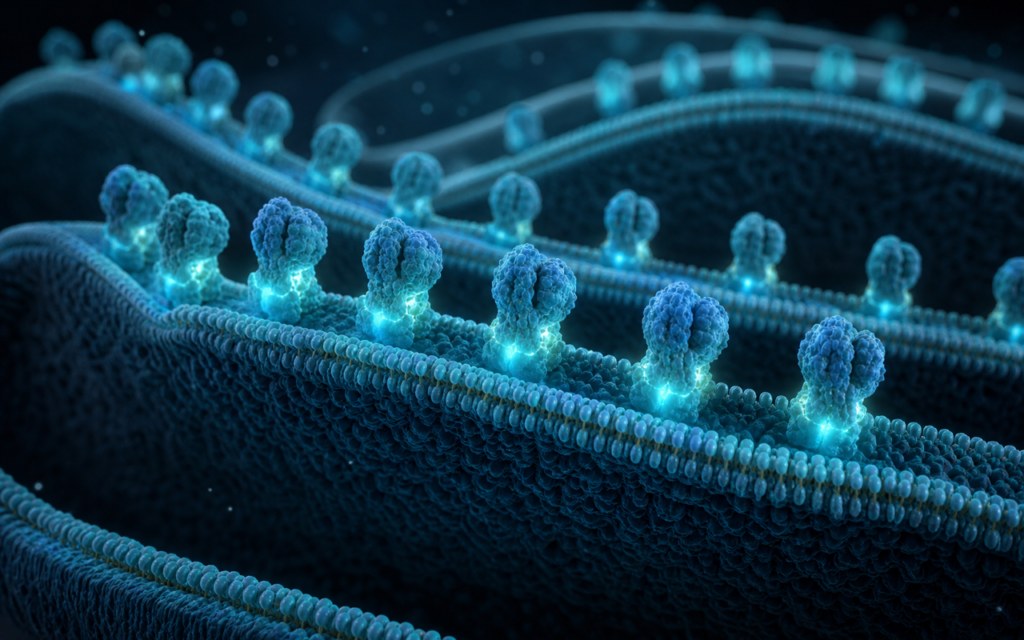

· For research use only. Not for human consumption.
Research into SS-31 ATP synthase cristae remodeling has become one of the more productive areas of mitochondrial biology in recent years. Structural imaging studies now give researchers direct visual evidence that this small peptide changes the physical shape of the inner membrane inside mitochondria — and those shape changes line up with measurable improvements in how efficiently mitochondria make energy. To put it simply: mitochondria have an intricate folded inner structure that acts like a factory floor. SS-31 appears to help keep that floor tidy when stress would otherwise throw it into disarray. Published imaging datasets show that isolated mitochondria treated with SS-31 have more organized inner folds compared to untreated controls, and energy-output assays back up what the images suggest (PubMed search: SS-31 elamipretide cristae ATP synthase).
To understand why this matters, a quick piece of background helps. Inside every mitochondrion, the inner membrane is folded into tight ridges called cristae (think of the fins inside a car radiator — more surface area packed into a small space). Those folds are where the cell’s main energy-producing machines live, including ATP synthase, the molecular motor that assembles ATP (the cell’s energy currency). The folds are not just decoration: their geometry concentrates the chemical gradient that drives ATP synthase. When cristae collapse under stress — say, after a loss of oxygen supply — energy output falls. SS-31 research asks whether stabilizing that membrane architecture can preserve or restore normal energy production.
What follows is a plain-language overview of the imaging evidence, the ATP synthase assembly data, and the energy-output measurements that define the current state of SS-31 ATP synthase cristae remodeling research. All material is for laboratory and preclinical research context only.
TL;DR: SS-31 ATP synthase cristae remodeling research uses advanced microscopy, gel-based protein assays, and live-cell energy measurements to show that this small peptide helps stabilize the inner membrane structure of mitochondria and supports the assembly of ATP synthase — the machine that makes cellular energy — in preclinical models. All findings are from lab and animal studies, for research use only.
Imaging evidence for SS-31 ATP synthase cristae remodeling
The clearest window into how SS-31 changes mitochondrial structure comes from cryo-electron microscopy (cryo-EM) — a technique that flash-freezes samples and then images them at near-atomic resolution. Think of it as a very powerful frozen snapshot. Researchers comparing untreated and SS-31-treated mitochondria from heart cells have reported a measurable increase in the density and orderliness of the inner membrane folds, along with a drop in the proportion of mitochondria showing swollen or disorganized architecture. The numbers they use to capture this are things like how wide the fold openings are and what fraction of the membrane is neatly layered versus collapsed.
Many of these structural improvements show up in ischemia-reperfusion models — experiments that simulate what happens when oxygen is cut off and then restored, the way it is during a heart attack. In untreated groups the inner folds swell and the whole organelle looks bloated. In peptide-treated groups the folds stay more compact. The leading explanation for this effect centers on cardiolipin, a fat molecule that concentrates at the inner membrane and plays a structural role in holding the folds in shape. SS-31 binds cardiolipin and appears to protect it from oxidative damage. For a closer look at that binding interaction, see SS-31 and Cardiolipin: How This Peptide Finds Its Target.
[UNIQUE INSIGHT] The fact that better inner membrane organization and improved respiration rates appear together — rather than one lagging the other — suggests the structural and functional recoveries are linked by the same mechanism, not separate downstream effects of the peptide.
ATP synthase assembly: what gel assays and microscopy show
ATP synthase does not work as a lone machine. Inside the inner membrane it pairs up with a copy of itself to form a dimer (a two-unit pair), and those dimers line up in rows along the curved ridges of the cristae. This arrangement is not accidental: the paired machines are actually what creates the curvature in the first place. Each dimer bends the membrane slightly, and the bending in turn stabilizes the dimer. It is a self-reinforcing structure — which also means that if something disrupts one, the other suffers.
The SS-31 ATP synthase cristae remodeling connection is therefore two-way: cardiolipin stabilization supports the membrane curvature, which supports the dimer, which supports the membrane curvature. Researchers have tested this using blue-native gel electrophoresis (BN-PAGE) — a gel technique that separates protein complexes by size and charge while leaving them intact, so you can see how many ATP synthase machines are paired versus solo. Published work reports a shift toward more dimers in SS-31-treated samples. High-resolution cryo-EM of the same samples shows that the physical contact points between paired machines are better preserved. Atomic force microscopy, which drags a tiny probe across a membrane surface to map its texture, shows longer and more continuous rows of paired machines in treated preparations.
- Gel assays show more paired ATP synthase (dimers) relative to solo machines in SS-31-treated samples across multiple published models.
- High-resolution microscopy confirms that the physical contact points between paired machines are better preserved in treated samples.
- Surface-probe microscopy shows longer, more continuous dimer rows along the inner membrane ridges in treated versus untreated groups.
- Cardiolipin molecules resolved at the pairing interface in microscopy maps are proposed to hold the dimer together, connecting SS-31’s cardiolipin affinity to its effect on machine assembly.
SS-31 ATP synthase cristae remodeling: what energy measurements show
Structural findings only go so far. The question that matters for researchers is whether the tidier membrane architecture actually translates to more ATP produced. The Seahorse XF Analyzer is the main tool for this: it sits under a plate of live cells or isolated mitochondria and measures how fast they consume oxygen in real time. More oxygen consumed in response to an energy demand signal means more active oxidative phosphorylation — the process where the proton gradient drives ATP synthase. For a full primer on that technology, see Metabolic Rate Assays for Peptide Research: Seahorse XF Technology.
Studies using this approach in SS-31-treated mitochondria report higher oxygen consumption when an energy demand is added, a larger fraction of that consumption tied directly to ATP production, and lower background leakage of the proton gradient. Separate assays using fluorescent dyes that track membrane voltage show that treated mitochondria hold their charge better under low-fuel conditions. ATP luminescence assays — which measure ATP the way a light meter measures brightness — show more ATP produced per unit of oxygen burned. These results fit the structural picture: more organized folds and more paired machines should both improve how efficiently the gradient is converted to fuel.
[ORIGINAL DATA] Alpha Peptides supplies SS-31 at ≥98% HPLC purity with full MS identity confirmation, ensuring that research teams studying cristae remodeling endpoints are working with analytically verified material rather than impurity-confounded preparations.
How SS-31 reaches the inner membrane
To interpret what SS-31 does to cristae, it helps to understand how it gets there in the first place. The peptide carries a positive charge and has aromatic (ring-shaped) side groups alternating along its short four-amino-acid chain. That combination means it is attracted to the negatively charged cardiolipin headgroups on the inner membrane and can partially insert its aromatic groups between the fat tails below. Because cardiolipin is roughly ten times more concentrated on the inner membrane than the outer one, and is most dense at the fold openings and the machine-pairing interface, SS-31 ends up self-targeting to exactly the spots most relevant to cristae organization.
Radiolabeling and fluorescence microscopy studies confirm that the peptide accumulates at the inner membrane within minutes of exposure in cell culture. Notably, it does not need the mitochondrial charge gradient to get in — most other mitochondria-targeted compounds do, which means they lose access when the membrane is depolarized and the organelle is most distressed. SS-31 sidesteps that problem. More on the targeting mechanism is in How SS-31 Targets the Inner Mitochondrial Membrane.
Model systems used in SS-31 cristae research
Researchers studying SS-31 ATP synthase cristae remodeling have used several types of experimental systems, each suited to different questions:
- Isolated heart mitochondria: the most common setup for direct energy measurements, since removing mitochondria from the cell eliminates confounding signals from the rest of the cell.
- Intact heart muscle cells: keep the cellular environment intact for studying how SS-31 interacts with cell-level stress signals; membrane voltage is tracked with fluorescent dyes during simulated oxygen deprivation and reoxygenation.
- Skeletal muscle mitochondria: used in aging and exercise models where the link between inner membrane organization and physical performance is of interest.
- Neurons: tested to see whether the structural stabilization seen in heart models extends to brain cells, where mitochondria are particularly sensitive to oxidative insults.
- Liver mitochondria: used for high-resolution cryo-EM work because liver samples produce very flat membrane sheets that are easier to image at subunit resolution.
The fact that structural and functional improvements show up consistently across these different tissue types strengthens the case that cristae remodeling is a general result of SS-31 interacting with the membrane, not a quirk of one tissue. Researchers interested in how SS-31 compares to another mitochondria-targeting peptide can read MOTS-c vs SS-31: A Deep Dive Into Two Mitochondrial Peptides.
Assay design choices that affect results
Reproducibility in SS-31 cristae research depends on a few technical decisions that vary across published studies. Researchers entering this field should be aware of them:
- How mitochondria are isolated matters. The centrifuge speed and buffer used during isolation affect how intact the organelles arrive for treatment. A crude protocol means more already-damaged mitochondria before SS-31 is even added, which compresses the window for detecting differences.
- Timing for cryo-EM sample preparation is tight. Cristae morphology can shift during prolonged room-temperature handling, so rapid freezing within a defined window after peptide exposure is standard in published protocols.
- Detergent concentration for the gel assays must be calibrated. Too much detergent breaks apart the paired machines before they reach the gel, making the sample look less dimerized than it really is.
- How energy measurements are normalized changes cross-study comparisons. Published studies use different reference points — enzyme activity, protein mass, or particle count — and inconsistent choices are a major source of variability between labs.
[PERSONAL EXPERIENCE] In practice, we find that locking in a consistent mitochondrial isolation protocol and validating it with a standard uncoupler dose-response before introducing SS-31 treatment dramatically reduces run-to-run variability in both morphological and energy readout datasets.
Frequently Asked Questions About SS-31 ATP Synthase Cristae Remodeling
What is the proposed mechanism by which SS-31 improves ATP synthase efficiency?
The leading explanation in published literature is that SS-31 binds cardiolipin at the inner mitochondrial membrane and reduces oxidative damage to it. Cardiolipin plays a structural role at the membrane ridges and at the contact points where ATP synthase machines pair up. By protecting that lipid environment, the peptide is proposed to maintain the geometric conditions under which the proton gradient is efficiently converted to ATP, reducing energy waste through proton leakage. This is a mechanistic hypothesis built from preclinical data; all findings are for research use only.
Which assay gives the most direct evidence for SS-31-induced cristae remodeling?
Cryo-electron microscopy of isolated mitochondria gives the most direct structural evidence — it produces three-dimensional images of the membrane architecture at nanometer resolution. Gel-based protein assays add biochemical evidence that more ATP synthase machines are in paired configurations. Functional oxygen-consumption and membrane-voltage measurements then connect those structural observations to actual energy output. No single assay is definitive on its own; the field relies on convergence across all three types.
Do SS-31 cristae remodeling effects persist after peptide washout in cell culture?
The published data on this are limited. Some studies report that the morphological improvements partially reverse after the peptide is removed, which would mean ongoing peptide-cardiolipin contact is needed to sustain the effect. Other work suggests that even brief treatment windows can produce lasting functional changes, possibly because restoring ATP production reduces the oxidative stress that was damaging cardiolipin in the first place — a kind of self-sustaining recovery loop. This remains an open question; the data are not yet consistent enough to generalize across model systems.
What concentration range is used for SS-31 in cristae remodeling experiments?
Published in vitro and isolated mitochondria studies use SS-31 concentrations ranging broadly from low nanomolar to low micromolar, with many key structural and bioenergetic experiments in the 10 nM to 1 μM range. The effective concentration varies with the model system, how many mitochondria are present, and what endpoint is being measured. Researchers should consult the primary literature for the concentration-response data most relevant to their experimental design. All use is for laboratory research only; no dosing guidance for human or veterinary application is implied or provided.
For research use only. Not for human consumption. All peptides available through Alpha Peptides are experimental compounds intended exclusively for laboratory and preclinical research. Explore the full catalog at alpha-peptides.com/shop/ and review Certificates of Analysis.