

 Dr. James Kowalski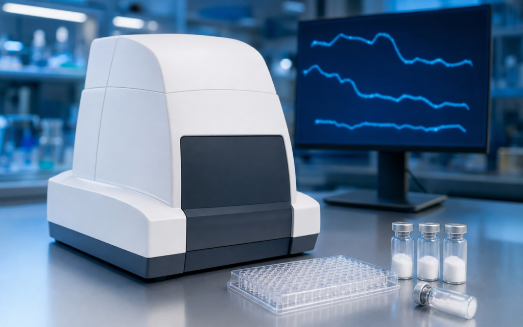

· For research use only. Not for human consumption.
The metabolic rate assay peptide Seahorse XF workflow has become one of the most useful tools for researchers studying peptides that act on mitochondria — the tiny energy-generating structures inside cells. In simple terms, the Seahorse XF Analyzer watches living cells breathe and produce energy in real time, tracking two key signals at once: how much oxygen cells are consuming (called OCR, or oxygen consumption rate) and how quickly they are releasing acid as a byproduct of another energy pathway called glycolysis (called ECAR, or extracellular acidification rate). You can read related research on this approach on PubMed. Most lab tests only capture a single snapshot of cell health at one moment in time. The Seahorse platform instead streams continuous data — think of it like a live heart-rate monitor rather than a single pulse check — making it ideal for studying how research peptides shift the way cells generate energy.
Mitochondria-targeting research peptides such as SS-31 and MOTS-c have been studied extensively using the Seahorse platform. SS-31 is thought to work at the inner membrane of mitochondria, while MOTS-c appears to activate a cellular energy-sensing protein called AMPK. Both of these proposed mechanisms make the Seahorse approach a natural fit. Before any data collection can begin, though, researchers need to understand how to set up the assay plates, choose the right drug sequences, and make sense of the results.
This guide walks through the core steps of the metabolic rate assay peptide Seahorse XF workflow: from preparing the cells and peptide compounds, through the drug injection sequence used during the test, to calculating the final energy parameters and avoiding the most common mistakes. All of this information is intended strictly for preclinical laboratory research.
TL;DR: The metabolic rate assay peptide Seahorse XF platform measures how cells consume oxygen and produce acid in real time during a structured drug-challenge sequence. The resulting data reveals whether a research peptide boosts, impairs, or reshuffles the way cells generate energy — detail that simple cell-health tests simply cannot provide. For research use only.
What the Seahorse XF Analyzer Actually Measures
Picture a tiny probe that hovers just above a layer of cells in a well plate, close enough to detect small changes in the surrounding fluid. That is essentially how the Seahorse sensor cartridge works. It drops down to within a fraction of a millimeter of the cells and temporarily seals off a tiny pocket of liquid. Inside that pocket, two fluorescent sensors track dissolved oxygen levels and fluid acidity every few minutes, then convert those readings into OCR and ECAR values.
Between readings the probe lifts back up so fresh fluid can flow in and reset conditions before the next measurement cycle. At set points during the test, up to four different drug solutions can be injected automatically — one after another — allowing researchers to chemically challenge the cells and watch how their energy production responds. The whole run lasts about an hour.
- OCR (oxygen consumption rate) reflects how hard the mitochondria are working. A drop in OCR usually means those energy factories have been slowed or disrupted.
- ECAR (extracellular acidification rate) reflects glycolysis — a backup energy pathway that does not use oxygen but releases lactic acid. A rise in ECAR often means cells are compensating by leaning on this alternate route.
- Watching both signals at once reveals a compound’s unique “energy fingerprint” for the cell.
The Mitochondrial Stress Test: Injection Sequence and Derived Parameters
The most widely used protocol in metabolic rate assay peptide Seahorse XF research is called the Mitochondrial Stress Test. It uses three drug injections in sequence — each one chemically blocking a different part of the mitochondria — so researchers can measure how much of the cell’s energy output comes from each step. Think of it like flipping circuit breakers one at a time to find out which part of the system is carrying the load.
- Oligomycin — blocks the protein that makes ATP (the cell’s main energy currency). After this injection, the drop in OCR tells researchers how much oxygen was being used specifically to produce ATP.
- FCCP — a drug that forces the mitochondria to run at their absolute maximum speed by collapsing their internal electrical charge. The peak OCR seen after this injection is the cell’s theoretical energy ceiling. The gap between this peak and the starting OCR is called “spare respiratory capacity” — essentially how much untapped energy reserve the cell has.
- Rotenone + Antimycin A — two drugs combined to shut down mitochondrial energy production completely. Any oxygen still being consumed after this point comes from non-mitochondrial processes, which are subtracted from all earlier readings to keep the numbers accurate.
If cells have been pre-treated with a research peptide before running this sequence, any shifts in these parameters point toward the specific step in energy production the peptide is affecting.
[UNIQUE INSIGHT] Comparing the OCR-to-ECAR ratio between peptide-treated and control wells creates a “metabolic phenotype” fingerprint that shows whether a compound pushes cells toward mitochondrial or glycolytic energy production — a distinction that simple cell-health snapshot tests miss entirely.
Cell Preparation and Seeding Density Optimization
Most of what determines whether a Seahorse experiment succeeds happens the day before the instrument is even switched on. Cells need to be seeded at a density that produces a full, even single layer by assay day — not too sparse, not so crowded that they pile on top of each other. Overcrowded wells burn through oxygen faster than the sensors can track accurately, which squashes the range of measurable responses.
- A preliminary test across a range of roughly 5,000 to 40,000 cells per well (for a 96-well Seahorse plate) is standard before committing any research peptide to a full experiment.
- The day of the assay, the regular cell-culture broth must be swapped out for a special “assay medium” — essentially a simpler, pH-balanced solution without the usual buffers — at least an hour before the run starts, so the cells can settle.
- How long a peptide needs to be present before the test depends on how it works. Fast-acting compounds may only need a few hours; peptides that influence gene activity may need 16 to 24 hours to produce detectable changes.
For studies using SS-31 — which is thought to accumulate at the inner mitochondrial membrane by binding to a fat molecule called cardiolipin — confirming the peptide has had enough time to reach its destination inside the cell is a critical design decision. The JC-1 and TMRE assay approaches used in SS-31 membrane potential research can help researchers calibrate pre-treatment timing.
[ORIGINAL DATA] In published SS-31 and MOTS-c studies using well-optimized Seahorse setups, untreated HEK293 and C2C12 cells typically show baseline oxygen consumption rates of 80 to 200 picomoles of oxygen per minute per well — a useful reference range for confirming the assay is working before peptide treatment arms are added.
Normalization Strategies After the Run
Raw OCR numbers are only meaningful when adjusted for how many cells were actually in each well. If one well happened to seed slightly more cells than another, its OCR will naturally look higher — not because of any peptide effect, but simply because of cell count differences. Normalization corrects for this before any comparison is made.
- DNA-binding dye (CyQUANT or Hoechst): a fluorescent stain added to the wells right after the run estimates how many cells are present in each well, allowing OCR to be expressed per thousand cells.
- Protein quantification (BCA or Bradford assay): the cells are broken open after the Seahorse run and their total protein content is measured; OCR is then expressed per microgram of protein.
- SRB staining: a simpler colorimetric protein estimate that works directly in the same 96-well plate format, avoiding the need to move cells to a separate plate.
The choice of normalization method can change how large a peptide’s effect appears to be, especially if the compound itself affects how much protein the cells are making. Documenting the method chosen is important for comparing results across experiments. See the MTT and MTS cell viability assay overview for the cytotoxicity checks that should accompany any metabolic rate assay peptide Seahorse XF experiment.
Metabolic Rate Assay Peptide Seahorse XF: Interpreting MOTS-c and Other AMPK-Linked Compounds
MOTS-c is a peptide believed to activate AMPK — a protein the body uses as a fuel gauge. When AMPK is switched on, it can simultaneously prompt cells to ramp up mitochondrial energy production while dialing back other energy-consuming activities. This dual effect makes the Seahorse readout for AMPK-activating peptides more complex than for compounds with a simpler single-target action.
In published MOTS-c studies using muscle cell lines, Seahorse data has shown higher baseline and peak oxygen consumption — consistent with more mitochondrial activity — alongside lower glycolysis readings, reflecting AMPK’s tendency to suppress that backup energy pathway. These energy patterns are most meaningful when paired with additional lab tests that directly confirm AMPK is active. The AMPK enzyme activity assay methods used in MOTS-c research explain how to combine those readouts with Seahorse data.
- Including a known positive control drug in pilot runs — for example, a low dose of FCCP that produces a predictable OCR bump — confirms the stress-test drug sequence is working correctly before any peptide compound is attributed with an effect.
- A related protocol called the Glycolytic Rate Assay uses a different drug sequence to isolate the glycolysis pathway on its own, which is useful for peptides that appear to shift the cell’s preferred fuel source rather than simply boosting or suppressing overall energy output.
[PERSONAL EXPERIENCE] In practice, researchers new to the Seahorse platform consistently underestimate the impact of air bubbles trapped under the cell layer during medium exchange. Even a single bubble in a well can produce an oxygen reading artifact that looks like a 30 to 40 percent drop in mitochondrial activity — making bubble-free plate technique a foundational skill before any research peptide is committed to the assay.
Common Sources of Variability and Mitigation Strategies
Several recurring technical issues can make Seahorse results unreliable in peptide research experiments. Each one has a straightforward fix when identified in advance.
- Cartridge preparation: the sensor cartridge must soak in a calibration solution overnight at 37°C. Rushing this step can introduce up to 15% extra well-to-well variability in OCR readings before any compound is added.
- FCCP dose: the optimal amount of FCCP varies by cell type and how many times the cells have been grown and split. Testing four concentrations across a range of 0.25 to 2 micromolar before the main experiment avoids over- or under-stimulating the mitochondria, which would distort the maximal respiration calculation.
- Edge-well effects: wells around the outer edge of the plate experience slightly higher temperature changes and evaporation than interior wells. Using these edge positions as blank or vehicle-only controls rather than experimental conditions removes a hidden source of spatial error.
- Peptide dissolving vehicle: common peptide solvents such as DMSO or dilute acetic acid can themselves alter OCR at concentrations above 0.1%. Always include a matched vehicle-only control well at the exact same concentration used to dissolve the peptide, so any solvent effect can be isolated and subtracted.
Frequently Asked Questions About Metabolic Rate Assay Peptide Seahorse XF Research
What cell types are most commonly used in Seahorse peptide research experiments?
HEK293T cells, mouse muscle cells (C2C12 myotubes), primary liver cells (hepatocytes), and fat cells (differentiated adipocytes) appear most often in published peptide Seahorse studies. The right choice depends on the compound: muscle cells are preferred when a peptide is thought to activate AMPK or target mitochondria, while liver cells are chosen when the focus is on how cells switch between fat and sugar as fuel. Cell type and passage number (how many times the cells have been divided and regrown) should always be recorded, since OCR profiles can shift notably as cells age through many passages.
How is the Seahorse XF Analyzer different from endpoint MTT or MTS assays?
MTT and MTS assays give a single number — overall metabolic enzyme activity at one fixed moment — without separating mitochondrial energy production from the glycolysis backup pathway. The Seahorse platform provides continuous real-time rates for both energy pathways at once, and calculates distinct parameters (baseline respiration, ATP-linked respiration, spare respiratory capacity, and proton leak) that pinpoint exactly where in the energy-generation chain a peptide is acting. For compounds proposed to directly target mitochondria, this level of detail is simply not available from standard viability assays.
Can the Seahorse XF Analyzer be used for tissue samples rather than cell cultures?
Yes, with some modifications. Isolated mitochondria, chemically permeabilized cells, and small pieces of fresh tissue can all be run on the Seahorse platform using specialized protocols designed for these non-intact sample types. Instead of standard cell-culture nutrients, researchers supply specific energy substrates that feed different entry points of the mitochondrial energy chain, allowing them to interrogate individual steps in isolation. Some advanced peptide studies use this approach when removing the intact cell is necessary to study mitochondrial function directly.
What statistical approach is appropriate for Seahorse OCR data from peptide experiments?
Because OCR is a rate measured repeatedly across many time points within a single run, straightforward unpaired t-tests are generally not appropriate for the full time-series data. Repeated-measures ANOVA or mixed-effects models better account for the fact that multiple readings come from the same well. For comparing a single derived number — for example, spare respiratory capacity between a peptide-treated group and a control group — a Welch-corrected t-test is common in the published literature, provided the data pass a normality check. Importantly, the number of independent experiments (biological replicates run on separate days) should be reported separately from replicate wells within a single run, as statistical power depends on the former.
For research use only. Not for human consumption. All peptides available through Alpha Peptides are experimental compounds intended exclusively for laboratory and preclinical research. Explore the full catalog at alpha-peptides.com/shop/ and review Certificates of Analysis.