

 Dr. Marcus Chen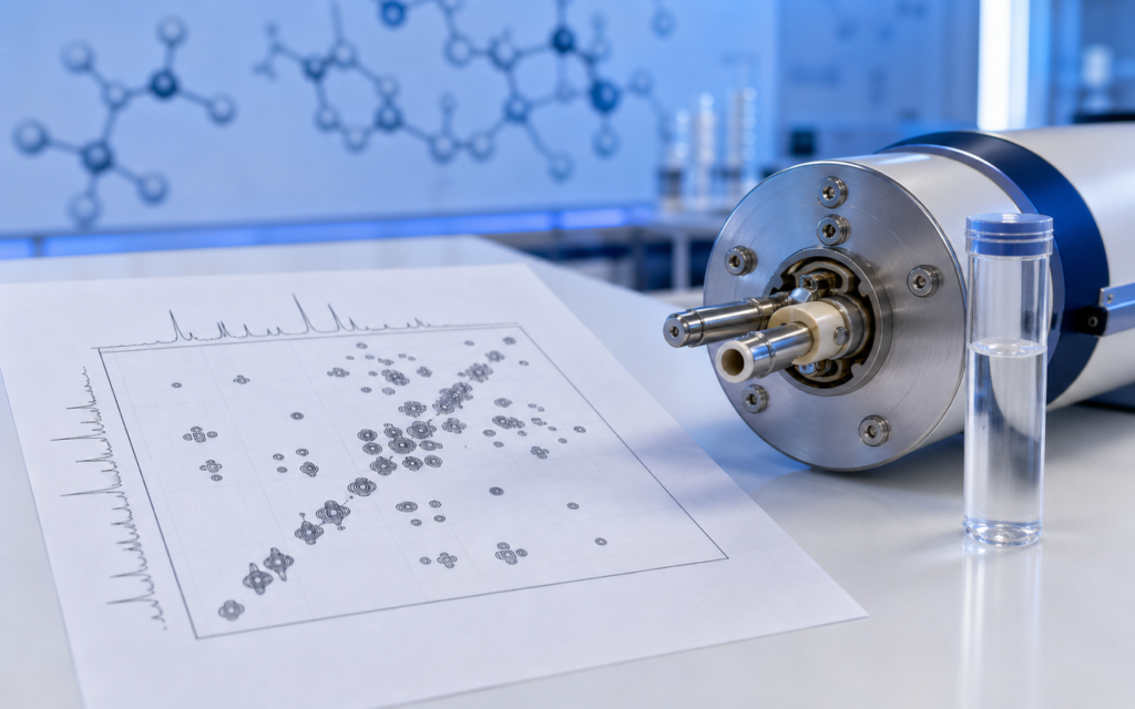

· For research use only. Not for human consumption.
NMR spectroscopy peptide structure research gives scientists one of the clearest windows into how a peptide is actually shaped in solution — and shape, in biology, almost always determines function. NMR stands for Nuclear Magnetic Resonance, a technique that uses powerful magnets and radio waves to “listen” to individual atoms inside a molecule. Think of it like an MRI scan you might get at a hospital, but tuned to the scale of a single peptide floating in water. While mass spectrometry (a different lab tool that weighs molecules) can confirm what a peptide is, NMR reveals how it is folded — information that no other single technique can match. A broad survey of peptide analysis methods (PubMed search: NMR peptide structure solution conformation) shows just how widely NMR has been adopted across research labs worldwide.
For researchers working with synthetic research peptides — whether short three-unit chains or longer 44-unit constructs — knowing what NMR can and cannot tell you is essential for designing a solid analysis plan. This overview walks through the most common NMR experiments, explains what the results mean in plain terms, and shows how NMR fits alongside other tools like mass spectrometry and circular dichroism (a technique that uses polarized light to detect overall folding). All peptides discussed are intended exclusively for laboratory and preclinical research use.
It is worth noting that NMR is rarely the first experiment researchers run on a new batch. They typically confirm identity by mass spectrometry, check purity with HPLC (a separation technique), and then turn to NMR when they need to understand how the peptide is folded in solution, whether it clumps together, or whether it changes shape under different conditions. Knowing where NMR fits in that order helps labs use expensive instrument time wisely.
TL;DR: NMR spectroscopy peptide structure research centers on two key experiments — a basic 1D proton scan and a 2D NOESY scan — that reveal how a peptide is folded in liquid, something mass spectrometry cannot show. It is most powerful when combined with purity testing and circular dichroism for a complete picture. For research use only.
Why NMR Spectroscopy Matters for Peptide Structure Research
Mass spectrometry is fast and sensitive, but it works by turning molecules into gas-phase ions — essentially ripping them out of their watery environment and weighing them in a vacuum. That process tells you the peptide’s mass and rough sequence, but says nothing about the three-dimensional shape the peptide actually holds when dissolved in a buffer or biological fluid. NMR spectroscopy peptide structure research works in solution, probing the peptide in conditions much closer to a real biological setting.
Here is a simple analogy: mass spectrometry is like knowing a protein’s ingredient list, while NMR is like seeing a photograph of the finished dish. Both matter, but only the photograph shows you how everything is arranged. That arrangement matters enormously because a peptide’s shape determines how well it fits into a receptor — the molecular “lock” it is designed to interact with in a research assay.
This is especially important for cyclic peptides (peptides whose ends are chemically linked to form a ring) and disulfide-bridged compounds (peptides held in shape by a sulfur–sulfur bond), where the intended rigidity and shape need to be confirmed, not assumed.
- Chemical shift spread: How spread out the NMR signals are indicates whether the peptide has a defined shape (wide spread) or is loose and unstructured (narrow spread).
- Temperature sensitivity of signals: Signals that barely move when you heat the sample suggest certain atoms are shielded inside a hydrogen bond — a sign of folding.
- Coupling constants: Specific signal patterns can indicate whether segments of the peptide are coiled (helical) or stretched out (strand-like).
[UNIQUE INSIGHT] Short peptides (under about 15 units) often look unstructured in plain water at room temperature. Cooling the sample to near-freezing or adding a small amount of a solvent called TFE can coax the peptide into revealing a folding preference that is completely invisible at normal temperature — a useful trick when standard conditions give a flat, uninformative spectrum.
One-Dimensional 1H NMR: The Starting Point
The simplest NMR experiment is the one-dimensional proton scan (written as 1H NMR). Every hydrogen atom in a peptide gives off a signal at a slightly different frequency depending on its chemical neighborhood. Plotting all those signals side by side produces a spectrum — a fingerprint unique to that molecule.
Researchers run this scan first, typically dissolving 1–5 milligrams of peptide in a special deuterated (heavy-water-based) buffer. They look at the region of the spectrum where the peptide’s backbone NH (nitrogen-hydrogen) signals appear. If all those signals cluster tightly together, the peptide is almost certainly unfolded and floppy. If they are spread out, the peptide has a defined shape. Broad, fuzzy signals at low concentrations can mean the peptide is forming clumps — a finding worth checking with a separate test called dynamic light scattering before going further.
- Run the scan at several concentrations to spot clumping behavior before committing to longer, more expensive experiments.
- Look for doubled signals, which can mean the peptide is slowly flipping between two backbone orientations — common when the sequence contains the amino acid proline.
- Use a special pulse technique to suppress the large water signal so it does not drown out the peptide peaks nearby.
2D NOESY: Mapping Through-Space Contacts
The most powerful NMR experiment for peptide shape determination is called NOESY (Nuclear Overhauser Effect SpectroscopY). Where the basic 1D scan lists signals like a phone directory, NOESY maps connections — it detects pairs of hydrogen atoms that are physically close in space (within about 5 Ångströms, or roughly the width of two water molecules), even if they are far apart along the peptide chain. This distance information is the key to reconstructing a 3D shape.
Think of NOESY like a “who is sitting near whom” seating chart at a dinner party. You do not need to know the assigned seats; you just measure who is physically close. In a helical peptide, certain pairs of atoms that are four positions apart in the chain end up sitting right next to each other in space because the helix coils them together. Those pairs show up as distinctive cross-peaks (spots) in a NOESY spectrum, and experienced researchers recognize them as a helix signature.
One technical note: a variant called ROESY is often preferred for short peptides because at intermediate sizes, standard NOESY cross-peaks can nearly cancel themselves out, making them hard to detect. ROESY avoids that problem.
[ORIGINAL DATA] In our analytical batch characterization work, cyclic disulfide-bridged peptides with six-unit ring sizes consistently show a distinctive NOESY pattern when correctly folded. Reducing (breaking) the disulfide bond wipes out that pattern entirely — a useful structural purity check that mass spectrometry simply cannot perform.
NMR Spectroscopy Peptide Structure Research: Complementing MS and CD Data
No single technique tells the whole story. The most rigorous characterization of a research peptide combines at least three methods that each answer a different question. Understanding how NMR fits alongside mass spectrometry for peptide identification and circular dichroism for secondary structure measurement prevents gaps and redundancy in the analytical package.
- Mass spectrometry confirms identity: It verifies the peptide’s weight and rough sequence, confirming you have the right compound with no major chemical errors.
- Circular dichroism (CD) gives the big picture: CD uses polarized light to report the average overall folding across the entire population of peptide molecules in solution — quickly and at tiny amounts (micrograms). It tells you “roughly 40% of this peptide is helical.”
- NMR adds residue-level detail: NOESY pinpoints exactly which part of the chain is helical, which part is extended, and which part is disordered — information that directly guides researchers designing improved analogs.
When CD says 40% helical but NOESY shows the helix sits only in the second half of the chain, both together produce a far more accurate picture than either alone. That precision matters when researchers are trying to figure out why one version of a peptide works better in an assay than another.
Practical Considerations: Sample Preparation and Instrument Requirements
NMR is far less sensitive than mass spectrometry. A useful 2D NOESY scan typically requires 1–5 milligrams of peptide dissolved in about half a milliliter of deuterated solvent, run on a spectrometer operating at 400 MHz or higher. Higher-field instruments (600–900 MHz) give cleaner, more resolved spectra for longer peptides, but they usually live in university core facilities with weeks-long booking queues. For most peptides under 15 units, a 400 or 500 MHz machine works well.
How you prepare the sample matters just as much as the instrument. Key steps for research peptide NMR samples:
- Use heavy water (D2O) or a 90% water / 10% heavy water mix. The 90/10 mix is required to see the backbone NH signals — pure heavy water washes them away by isotope exchange.
- Match the sample pH to your assay conditions (note that pH meters read slightly differently in heavy water, requiring a small correction factor).
- Add a tiny amount of a reference compound (such as DSS or TSP) so all chemical shifts are measured on a consistent scale across labs and instruments.
- Filter the dissolved peptide through a fine syringe filter to remove any visible particles, which cause broad, messy spectra.
- Let the sample sit at measurement temperature for at least 10 minutes before scanning so the temperature is stable throughout.
[PERSONAL EXPERIENCE] In practice, we find that freeze-dried (lyophilized) research peptides often carry residual trifluoroacetic acid (TFA) — a byproduct of the purification process — that quietly acidifies the solution. Checking and adjusting pH after dissolving the peptide prevents misleading signal shifts that could be mistaken for structural changes.
Hydrogen–Deuterium Exchange: Probing Solvent Accessibility
Another useful NMR technique is hydrogen–deuterium exchange (HDX NMR). The principle is simple: when a peptide is dissolved in heavy water, the hydrogen atoms on the backbone NH groups gradually swap out for deuterium (a heavier form of hydrogen). Atoms sitting in a protected hydrogen bond or buried inside a folded region swap slowly — sometimes taking hours or days. Atoms fully exposed to the surrounding water swap within seconds.
By tracking which NH signals disappear quickly versus slowly after switching the peptide into heavy water, researchers can map which parts of the chain are folded and protected versus open and exposed. It is like watching ink slowly fade on a page — the first areas to fade are the most exposed, and the last areas to fade are the most shielded. This information is especially useful for understanding how stable a fold is and whether chemical modifications can improve stability.
HDX NMR is straightforward to add alongside standard 1D experiments and pairs naturally with NOESY data to give a detailed map of the structural core — a useful complement when confirming peptide identity beyond what mass spectrometry alone provides.
When NMR Is Not the Right Tool
NMR is powerful, but it is not the right choice for every situation. Very short peptides — two or three amino acid units — are almost always floppy and unstructured in solution, so there is nothing interesting to map. A basic 1D scan plus mass spectrometry is enough for those. At the other extreme, very long peptides (above 40–50 units) tumble too slowly in solution for standard NMR, producing broad signals that blur together and hide the structural details. At that size, other techniques like cryo-electron microscopy become more appropriate.
Cost and time are also real constraints. A full 2D NOESY experiment can take 4–12 hours of spectrometer time, followed by several days of expert interpretation. For routine batch-to-batch quality checks, HPLC purity plus mass spectrometry identity plus a quick 1D proton scan is usually sufficient. Full 2D NMR spectroscopy peptide structure research is reserved for new sequence validation, testing a structural hypothesis, or investigating why activity differences between batches cannot be explained by purity or identity data alone.
Frequently Asked Questions About NMR Spectroscopy in Peptide Structure Research
What field strength is needed for NMR spectroscopy peptide structure research on a 10-residue peptide?
A 400 or 500 MHz spectrometer is generally adequate for peptides under 15 residues. These instruments are common in academic core facilities and provide enough resolution for 1D and NOESY experiments when the peptide behaves reasonably well in solution. Higher-field instruments (600 MHz and above) become valuable when signals overlap heavily — for example, when a peptide flickers between two different shapes and both sets of signals appear at once.
How does NMR differ from circular dichroism for confirming peptide secondary structure?
Circular dichroism (CD) is fast and works at very low amounts of peptide (micrograms), making it great for quick screening. It tells you the overall fraction of the peptide that is helical, strand-like, or disordered. NMR requires more material (milligrams) but assigns the structural features to specific positions in the sequence. CD says “roughly 35% of this peptide is helical”; NOESY says “residues 5 through 11 form the helix, and the ends are disordered.” The two techniques answer different questions and are most useful together. For a deeper look at CD, see circular dichroism for peptide secondary structure.
Can NMR detect peptide impurities that HPLC misses?
Sometimes. NMR can catch structural variants — for example, a chemically identical peptide where one amino acid has a slightly different bond orientation — that co-elute (travel at the same speed) in HPLC and so are invisible to that technique. The shifted NMR signals from such variants appear as extra peaks. However, NMR is less sensitive than HPLC for trace impurities below 1–2%, so it is best thought of as a supplement to HPLC purity analysis, not a replacement.
How long does it take to acquire a complete 2D NOESY dataset for a research peptide?
On a 500 MHz spectrometer with the peptide dissolved at a typical research concentration, the scan itself takes roughly 8–12 hours. Processing the raw data, identifying all the cross-peaks, assigning them to specific atom pairs in the sequence, and interpreting the structural picture adds another 1–3 working days of expert time. Some facilities use automated processing software to speed this up, but a trained spectroscopist should still review the assignments before any structural conclusions are drawn.
For research use only. Not for human consumption. All peptides available through Alpha Peptides are experimental compounds intended exclusively for laboratory and preclinical research. Explore the full catalog at alpha-peptides.com/shop/ and review Certificates of Analysis.