

 Dr. James Kowalski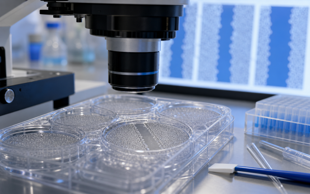

· For research use only. Not for human consumption.
Peptide wound closure scratch assay research gives scientists a simple, affordable way to watch cells close a gap — and to figure out whether a peptide is helping that happen. The scratch wound assay (often called the scratch test) works exactly as it sounds: a researcher drags a fine tip across a dish of cells, creating a bare stripe, then watches to see how quickly the cells move back in. Paired with a second method called the Boyden chamber test, researchers can not only measure closure speed but also tell why it is happening. This guide breaks both methods down in plain terms, so you can follow the science even without a lab background. (PubMed: scratch wound assay cell migration)
Why run two tests instead of one? Because the scratch test shows the end result — did the gap close? — without telling you the reason. Cells can fill a gap by moving into it (migration) or by dividing and multiplying near the edge (proliferation). A separate migration-only test, the Boyden chamber (think of it as a mini obstacle course cells must travel through), separates those two explanations. A peptide that closes wounds but fails the migration-only test is probably working by speeding up cell division, not movement — a meaningful difference for research purposes. Understanding this distinction is fundamental to cell-based assays for peptide research.
This guide covers both protocols step by step, including setup requirements, important controls, imaging tips, and common mistakes. All content is for preclinical laboratory research only. For research use only. Not for human consumption.
TL;DR: Peptide wound closure scratch assay research combines the scratch wound method (2-D gap closure) with the Boyden chamber migration assay (directed chemotaxis) to separate proliferative from migratory peptide effects. Rigorous controls, consistent imaging intervals, and validated image analysis are the difference between publishable data and confounded results. For research use only.
How the In Vitro Scratch Wound Assay Works
Picture a thick lawn of cells covering the bottom of a lab dish — so dense they are all touching. A researcher drags a fine pipette tip or scraper across the dish in a straight line, clearing a tidy lane through the cells. That bare lane is the “wound.” Over the next several hours, a camera captures images at regular intervals. Researchers then measure how much of that lane has been filled back in by the returning cells — expressed as a percentage of the original gap. That number is the main result: percent wound closure.
For peptide wound closure scratch assay research to give clean, trustworthy data, a few things must be set up carefully before the scratch is made:
- A fully covered dish: Cells should cover essentially the entire surface (95–100%) before scratching. Patchy coverage creates uneven wound edges that make measurements messy and inconsistent.
- Reducing the growth signal briefly: Switching cells to a low-nutrient medium for a few hours before scratching settles the cells into a resting state. This keeps background cell division low so any growth seen later can be attributed to the peptide treatment.
- A consistent scratch width: The gap should be a uniform size — roughly the width of a thin marker line (400–800 micrometers). Automated tools that scratch all wells of a plate simultaneously make this much more reproducible than scratching by hand.
- Cleaning up afterward: Once the scratch is made, the medium is replaced. This removes floating debris and loose cells that could skew gap measurements.
The peptide being studied is added right after this cleanup step. A “vehicle control” — the same liquid the peptide was dissolved in, but without the peptide — is always run alongside as a comparison. See the lyophilized peptide reconstitution protocol for the correct way to prepare peptides before adding them to cells.
Separating Cell Movement from Cell Division
The biggest weakness of the scratch test on its own is that cells can fill the gap two ways: by moving (migration) or by dividing and multiplying (proliferation). If a peptide accelerates closure, you need to know which of those it is driving. That is where mitomycin-C (MMC) comes in.
MMC is a chemical that pauses cell division without immediately harming the cells. Treating cells with MMC before the scratch means any gap closure that happens afterward can only be due to movement, not multiplication. Standard treatment is a 2-hour soak at a set concentration, followed by a thorough rinse. After that, scientists proceed with the scratch as normal.
Comparing MMC-treated wells to untreated wells reveals the story clearly. If the peptide closes the wound in both conditions, movement is the main driver. If closure drops significantly in the MMC-treated wells, cell division is playing a big role.
[UNIQUE INSIGHT] Researchers who include both MMC-treated and untreated wells across a range of peptide concentrations can estimate how much of the closure effect comes from movement versus division at each dose — a more informative read-out than closure rate alone, and one that directly informs whether a peptide candidate warrants further study of cell movement machinery versus growth signaling.
Boyden Chamber Migration Protocol for Peptide Research
If the scratch test is a race across an open field, the Boyden chamber test is an obstacle course. Cells are placed on one side of a thin membrane (like a very fine mesh screen) punctured with tiny holes just large enough for cells to squeeze through. A chemical signal that attracts cells is placed on the other side. After several hours, researchers count how many cells made it through — those are the ones that actively moved toward the signal.
Key setup details for peptide wound closure scratch assay research using the Boyden format:
- Hole size in the membrane: 8 micrometers wide for most common cell types — big enough for a cell to squeeze through, but only if it actively tries.
- Membrane coating: The mesh can be pre-coated with proteins that mimic the natural scaffolding cells move through in the body, making the test more realistic.
- Cells on top, signal below: Cells are placed in the upper chamber in a nutrient-poor medium. The chemical signal (and the peptide being tested) goes in the lower chamber. Cells that sense the signal migrate down through the mesh.
- Counting the travelers: After the incubation period, the top of the membrane is wiped clean, and only the cells that crossed through are stained and counted under a microscope. Five different spots on each membrane are counted and averaged.
For researchers working with peptides like GHK-Cu — which has a notable body of preclinical literature on skin cell activation and tissue remodeling — the Boyden chamber provides a clean answer to whether the observed effects include genuine directed cell movement. Related background is covered in the post on GHK-Cu and fibroblast research studies.
[ORIGINAL DATA] In fibroblast migration experiments using peptide wound closure scratch assay research designs, the variability in scratch width at the starting point commonly falls below 8% when using an automated 96-well scratching tool, versus up to 22% with manual pipette-tip scratching — underscoring that instrument-assisted wounding is the single highest-leverage improvement most labs can make.
Image Acquisition and Analysis Methodology
Getting good data from the scratch test depends just as much on how images are captured as on the biology itself. Inconsistent lighting, a shift in camera position, or uneven timing between photos can introduce errors that no math can fully fix afterward. These straightforward conventions keep imaging clean across any peptide wound closure scratch assay research series:
- Mark your spots: Use software or physical reference marks to return the camera to the exact same position in each well at every time point. Losing track of position is the most common reason scratch assay data becomes unusable.
- Photograph regularly: Take a photo immediately after cleaning up post-scratch, then every 4–6 hours for up to 24–48 hours. At least four time points are needed to track how quickly the gap closes over time rather than just measuring a single end-point snapshot.
- Analysis tools: Free software like ImageJ (with the MRI Wound Healing plug-in) is widely used to measure gap area from each image. Automated live-imaging systems such as IncuCyte can photograph and measure continuously without human intervention, eliminating manual measurement error entirely.
- Normalize the numbers: Express results as: (starting gap size − gap size at each time point) ÷ starting gap size × 100. This percentage accounts for any starting differences in scratch width between wells.
For Boyden chamber images, capture all photos at the same exposure and apply the same brightness threshold before counting cells. Labeling cells with a fluorescent dye before the assay allows automated counting software to do the work — more consistent than manually counting stained cells afterward.
Peptide Wound Closure Scratch Assay Research: Controls and Common Pitfalls
Even a well-planned study can produce misleading results without the right comparison groups. A complete set of controls for peptide wound closure scratch assay research includes:
- Vehicle control: The same liquid the peptide was dissolved in, at the same volume, without the peptide. This ensures any observed effect is coming from the peptide and not its carrier liquid.
- Positive migration control: A substance known to trigger strong cell movement (such as EGF, a well-studied growth signal) is run alongside to confirm the assay is working and to give a benchmark for comparison.
- Division-blocked control: The MMC-treated wells described earlier, to separate movement from multiplication.
- Cell health check: Run a viability test on parallel wells at the same peptide concentrations. A peptide that appears to slow wound closure might just be harming the cells — a very different conclusion.
- True biological replicates: Ideally, at least three separate experiments using cells from different growth passages, each with triplicate wells. Running triplicate wells from a single experiment is technical repetition, not true biological replication.
[PERSONAL EXPERIENCE] In practice, we have found that skipping the photo immediately after the scratch — jumping straight to a 6-hour image — is a recurring mistake in early-stage labs. Without that first baseline measurement, there is no way to calculate percent closure accurately, and the entire dataset becomes difficult to interpret.
Connecting Scratch and Boyden Data to Peptide Mechanism
Running both assays side by side pays off most during data interpretation. Here is a plain-language breakdown of what each combination of results suggests:
- Scratch closure faster, more cells crossed the membrane: The peptide appears to genuinely promote cell movement. Follow-up studies would look at the internal machinery cells use to crawl — the structural proteins and signals that drive movement.
- Scratch closure faster, membrane crossings unchanged: Cell multiplication near the wound edge is likely the main driver, not movement. Follow-up would look at cell division markers.
- Scratch closure unchanged, more cells crossed the membrane: The peptide nudges cells to move in a directed way, but something may also be slowing net closure — a rare result, but one worth investigating carefully.
- Both results unchanged: No effect detected at these concentrations. Worth testing a broader range of doses, or confirming the peptide was active using a separate biological check.
Interpreting these results correctly also requires understanding what lab-dish data can and cannot tell us. Understanding these relationships requires a solid grounding in what in vitro versus in vivo data actually means for translational context. Scratch and Boyden results describe single-cell-type behavior in a dish — useful, but simplified. Findings should be confirmed with additional tests (such as 3-D cell cluster invasion models) before drawing firm conclusions about mechanism.
Frequently Asked Questions About Peptide Wound Closure Assay Research
What cell lines are most commonly used in peptide wound closure scratch assay research?
Human dermal fibroblasts (skin cells that produce structural proteins), HaCaT keratinocytes (a skin surface cell line), and NIH 3T3 mouse fibroblasts are the most common choices. They are well-studied, behave predictably, and have reliable published reference data for comparison. Primary cells taken directly from tissue are more realistic but harder to work with consistently. The right choice depends on the biological question: keratinocytes for studying the outer skin layer healing, fibroblasts for studying deeper tissue remodeling.
How do I account for peptide stability during a 24-hour scratch assay incubation?
Some research peptides can break down in cell culture medium over time, especially when the medium contains enzymes that cleave proteins. To address this: use reduced-enzyme conditions, refresh the peptide at each medium change, or test how much active peptide remains at different time points using analytical chemistry tools. Documenting what stability conditions were used is important so the experiment can be reproduced by others.
What is the difference between chemokinesis and chemotaxis in Boyden chamber assays?
Both words describe cell movement, but they differ in directionality. Chemotaxis is movement toward a concentration gradient — the cell is following a signal like a beacon. You test this by placing the peptide only in the lower chamber. Chemokinesis is a general boost to cell movement with no preferred direction — tested by putting equal amounts of the peptide in both the upper and lower chambers, eliminating any gradient. Knowing which one is occurring tells researchers whether the peptide is acting as a directional guide or simply making cells more restless overall.
Can I use the scratch assay for 3-D spheroid or organoid models?
The standard scratch test is designed for flat (2-D) cell layers and does not directly apply to 3-D cell clusters. For 3-D models, researchers instead grow compact cell spheroids and then transfer them onto a gel-like surface, watching how the cluster spreads outward over time — the 3-D equivalent of scratch closure. This approach is more technically demanding but better captures how cells behave in tissue-like environments with layers and structure.
For research use only. Not for human consumption. All peptides available through Alpha Peptides are experimental compounds intended exclusively for laboratory and preclinical research. Explore the full catalog at alpha-peptides.com/shop/ and review Certificates of Analysis.