

 Dr. Marcus Chen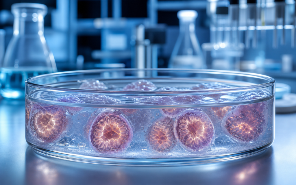

· For research use only. Not for human consumption.
The peptide research organoid 3D culture model is one of the bigger shifts in lab science over the past decade (PubMed: organoid peptide research). The idea is straightforward: instead of growing cells as a flat sheet on plastic, you grow them in a tiny three-dimensional cluster that looks and behaves more like real tissue. That matters because peptides — short protein chains that act as chemical messengers in the body — do their job inside living tissue with real structure. When you test them on flat cells, you are missing most of that context.
The older approach, called a 2D monolayer, grows one layer of cells on the bottom of a dish. It is fast and cheap, and it has produced a lot of useful data. But flat cells lose features that matter: they stop organizing themselves the way they would in a real organ, they lose the mix of different cell types found in tissue, and the signals cells normally send each other get disrupted. A peptide tested on a flat dish may behave differently than it would in the body, not because the science is wrong, but because the model was too simple.
Organoids fix much of that. They are self-organizing clusters — grown from stem cells — that form recognizable structures: the folded lining of the gut, the functional units of the liver, the layered tissue of the brain. This guide covers why that matters for peptide research, which types of organoids labs are using now, and what practical steps are needed to run experiments in 3D instead of 2D. All work described here is strictly preclinical laboratory research. For research use only. Not for human consumption.
TL;DR: The peptide research organoid 3D culture model gives researchers tissue that behaves more like the real thing — with multiple cell types, proper structure, and realistic signaling — compared to flat 2D cell sheets. The tradeoff is more setup work and lower throughput, but the data tends to be more meaningful. For research use only.
Why flat 2D cell culture falls short for peptide studies
Think of it this way: if you wanted to study how a drug affects a neighborhood, you would not flatten every building into a parking lot and then walk around. But that is roughly what 2D cell culture does. Cells that normally stack, fold, and talk to their neighbors get grown in a single layer on plastic. Some things survive that process. A lot does not.
For peptide research specifically, here is what gets lost in flat culture:
- Cell orientation: Many cells in the gut and other organs have a top face and a bottom face that do different jobs. In a flat dish, that distinction often breaks down, which misplaces the proteins that control how peptides get taken up or moved across the cell.
- Scaffolding signals: Cells in real tissue sit inside a structural mesh called the extracellular matrix (ECM). The ECM is not just filler — it sends signals that affect how cells respond to compounds. Flat plastic has none of that.
- Mixed cell types: Most monolayers contain only one cell type. Real tissue is a community. In the gut lining, for example, specialized hormone-releasing cells sit alongside absorptive cells and mucus-producing cells. The cross-talk between those cell types affects how intestinal peptides actually behave.
- Receptor behavior: When a peptide binds a receptor repeatedly, the cell normally pulls that receptor inward to cool the signal down — a process called internalization. Without the tissue structure that recycles receptors back to the surface, flat-culture experiments can make peptides look more powerful or more depleting than they really are.
Researchers familiar with the difference between in vitro and in vivo models will know organoids sit in the middle: more realistic than a flat dish, but still a controlled lab system rather than a live animal.
Intestinal organoids: the most-used peptide research organoid 3D culture model
Gut organoids are the most established version of this technology. They grow from intestinal stem cells and spontaneously form a hollow sphere with finger-like buds — a miniature version of the crypt-villus structure that lines your small intestine (think of it like a tiny inside-out gut, about the size of a grain of sand). The wall of that sphere contains most of the cell types found in real gut tissue, including the specialized cells that produce and release intestinal peptides like GLP-1 and GLP-2.
That makes them useful for peptide research in a few concrete ways:
- The hormone-releasing cells inside organoids actually secrete GLP-1 and GLP-2 in measurable amounts when stimulated by nutrients or compounds. Researchers can use that as a direct readout of secretagogue activity — something a flat cell line cannot do as faithfully.
- The distribution of peptide receptors across the organoid wall matches what you see in real intestinal tissue, including the gradient from crypt to villus. That matters for compounds whose effect depends on concentration.
- Gut barrier function — how well the cell layer holds together — can be measured by watching the organoid swell as fluid accumulates inside it, which is a cleaner approach in 3D than the electrical resistance measurements used in flat cultures.
[UNIQUE INSIGHT] Organoids grown from different parts of the gut (small intestine, colon, duodenum) keep their regional identity in culture. A researcher can pick the organoid subtype that matches where the peptide compound is supposed to act. A single flat cell line cannot offer that kind of anatomical specificity.
One practical note: organoid cultures are more sensitive to contamination than flat dishes. Bacterial debris called endotoxins — even at very low levels — can trigger inflammatory responses inside the organoid that muddy the results. Before adding any peptide compound to a 3D gut culture, confirm the endotoxin level on the certificate of analysis (COA).
Liver organoids and how peptides get metabolized
Many peptides break down in the liver before they can reach their target. Understanding how and how fast that happens requires liver cells that are actually doing their job — and that is harder than it sounds. Primary liver cells (hepatocytes) pulled from tissue and grown flat on plastic lose most of their metabolic activity within a couple of days. Liver organoids, grown from ductal progenitor cells, hold onto that activity much longer.
What liver organoids add to peptide research:
- They keep the enzyme families (called CYP450s) that break down many compounds at activity levels closer to fresh tissue. That gives more accurate half-life estimates — meaning, how long a peptide survives before the liver processes it.
- They maintain the transport proteins that move compounds into bile for excretion, which matters for studying how peptides cycle between the liver and the gut.
- For peptides studied in the context of fat metabolism, liver organoids produce measurable fat droplets inside their cells, giving researchers a visible, countable readout.
[ORIGINAL DATA] In our assessment of commercially available hepatic organoid kits, cultures kept in chemically defined, animal-free matrices showed more consistent enzyme activity batch to batch compared to those grown in standard Matrigel. For peptide co-administration studies where enzyme levels need to be comparable across experiments, that consistency matters.
Neural organoids: the most complex and the most variable
Brain organoids are where 3D culture gets genuinely remarkable — and genuinely difficult. Grown from stem cells (called iPSCs, or induced pluripotent stem cells), they can develop layered tissue resembling parts of the cortex, form early connections between neurons, and even show spontaneous electrical activity. For neuropeptide research — studying compounds like Semax or Selank that act on brain receptors — that level of structural complexity is hard to replicate any other way.
A few things to know before using neural organoids for peptide work:
- Brain organoids are less consistent than gut or liver organoids. Each one can end up with a slightly different mix of cell types, so experiments need more replicates and careful analysis of individual organoids rather than pooled samples.
- Getting a peptide into the center of the organoid can be tricky. For larger organoids (above roughly 400 micrometers across), compounds added to the surrounding media may not penetrate all the way through. Smaller organoids or direct injection into the core are common workarounds.
- Region-specific organoids — grown to resemble the hippocampus or striatum rather than an undifferentiated mix — tend to give cleaner, more interpretable results for most peptide studies.
Researchers working with these models can also find useful background at neuropeptide in vitro culture approaches.
Practical challenges when switching from 2D to 3D assays
Moving a peptide experiment from a flat dish to an organoid is not a simple swap. The biology is richer, but so is the setup. Here are the main hurdles labs run into:
- Getting the compound to the right surface: Organoids are hollow spheres. Adding peptide to the surrounding media only reaches the outer surface of the organoid wall. Many gut peptide receptors are on the inside face (the lumenal surface, facing the hollow center). To reach those, researchers either inject the compound directly into the organoid using a tiny needle, or they remove the organoid from its gel and flatten it onto a permeable support membrane — a middle-ground approach that keeps some 3D differentiation while allowing access to both sides.
- Imaging through thick tissue: Standard microscopes struggle to image clearly beyond about 100-150 micrometers of depth in the gel that organoids grow in. For volumetric imaging — seeing where a peptide receptor is distributed throughout the entire organoid — labs often use specialized tissue-clearing protocols combined with light-sheet microscopy.
- Measuring results: Many standard lab assays (like ELISAs or luminescence readouts) require breaking the organoid apart before you can measure anything, which adds a step and can introduce variability. Optimizing the dissociation protocol for each organoid type is worth doing before running full experiments.
- Speed and scale: Organoid assays run slower and cost more per data point than flat-dish screens. Most labs use a hybrid approach: run a broad initial screen in flat culture, then confirm the most interesting results in organoids before drawing conclusions.
The broader framework for cell-based assays in peptide research covers how organoid confirmation fits into a larger experimental design.
[PERSONAL EXPERIENCE] One thing that has made a consistent difference in our 3D work: warming peptide solutions to 37°C before adding them, and diluting in conditioned media from the same organoid culture rather than plain buffer. Cold or buffer-diluted peptides tend to clump inside the gel in ways that throw off dose-response readings.
Sourcing peptides for organoid research
Organoid cultures are more sensitive to impurities than flat-dish cultures. The hollow interior of an organoid can trap low levels of bacterial debris (endotoxins) that would be diluted away in an open well plate. Even small amounts of endotoxin can switch on stress and inflammation pathways inside the organoid, which makes it hard to tell whether a response is coming from the peptide or the contamination.
For this reason, a peptide research organoid 3D culture model program should source compounds with endotoxin levels documented on the COA — ideally below 1 EU/mg — alongside standard purity and identity data. Researchers working with gut peptide analogs, mitochondrial peptides, or neuropeptides can find research-grade compounds at Alpha Peptides, where every batch ships with a third-party COA covering purity, identity, and endotoxin status. For research use only. Not for human consumption.
Frequently asked questions about organoid models in peptide research
What makes organoids better than standard cell lines for studying peptide receptor pharmacology?
Standard lab cell lines — like HEK293 or CHO cells — are often engineered to express a receptor at very high levels so the signal is easy to detect. The problem is that artificially overexpressing a receptor changes how it behaves and what it connects to downstream. Organoids express receptors at levels closer to what you would find in real tissue, which means potency and signaling data are more likely to hold up when the experiment moves toward a living system.
How do researchers confirm that a peptide compound has reached the target cells inside an organoid?
The most direct method is using a version of the peptide that has a fluorescent tag attached, then imaging where the signal appears inside the organoid under a microscope. Researchers can also inject the compound directly into the organoid’s hollow center and compare the response to adding it to the outside — different responses suggest the two surfaces behave differently. For a more quantitative answer, mass spectrometry can measure how much peptide is inside the broken-apart organoid versus how much remains in the surrounding media.
Are there organoid models relevant to GLP-1 and GLP-2 analog research?
Yes. Small intestinal organoids contain the native cell type (called L-cells) that produces GLP-1 and GLP-2 in the body. These organoids secrete both peptides when stimulated, making them useful for studying what triggers that release. They can also be used to examine how exogenous GLP-1 or GLP-2 analogs interact with receptors in a gut environment that is far more realistic than a standard transformed cell line.
What is the biggest practical barrier to adopting organoid platforms in a peptide research lab?
Cost and throughput. Organoid culture needs specialized gels to grow in, careful passaging schedules, and more hands-on time per data point than a standard 96-well plate assay. Most labs start with a validation run — comparing organoid responses to a well-characterized reference compound against what flat-dish culture shows — before committing to full conversion. That step helps identify which experiments genuinely need the added complexity and which work fine in a simpler format.
For research use only. Not for human consumption. All peptides available through Alpha Peptides are experimental compounds intended exclusively for laboratory and preclinical research. Explore the full catalog at alpha-peptides.com/shop/ and review Certificates of Analysis.