

 Dr. Elena Vasquez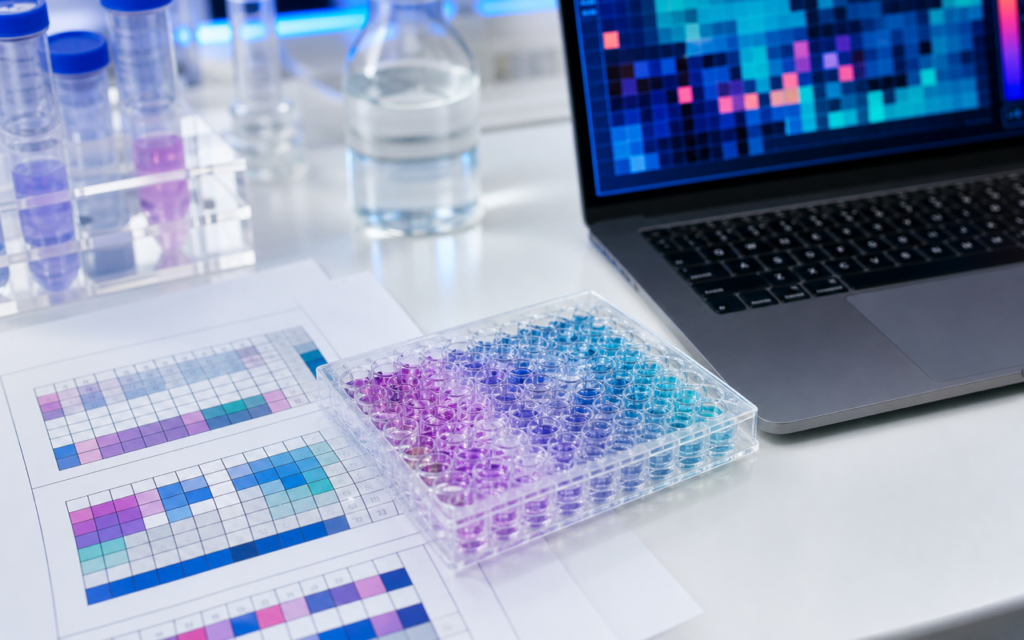

· For research use only. Not for human consumption.
Peptide library diversity design positional scanning is one of two main methods researchers use to figure out which amino acids — the building blocks of peptides — matter most for biological activity in a large mixed library. Think of it like this: instead of testing millions of individual combinations one by one, you group them cleverly and test the groups. One position in the sequence is held fixed while everything else varies. By seeing which fixed choice lights up the assay, you learn what the ideal building block is at that spot. Houghten and colleagues described this approach in the early 1990s (PubMed: positional scanning combinatorial libraries), and it has been a standard tool for finding active peptide leads ever since. The competing strategy, iterative deconvolution, reaches the same goal but works through multiple rounds of synthesis and testing rather than all at once.
Both methods face the same problem: a six-amino-acid peptide built from 20 possible amino acids has 64 million possible sequences. No lab can make and test each one. Mixture-based libraries solve this by pooling sequences together in a structured way and using assay readouts to point toward the best choices. The difference between positional scanning and iterative deconvolution is how they extract that information and how many rounds of lab work are needed.
For labs running peptide library screening for new bioactive sequences, picking the right strategy can mean the difference between finishing in one experiment or spending months on repeat synthesis cycles. This post explains how each approach works, where each one falls short, and how to choose between them.
TL;DR: Peptide library diversity design positional scanning reads all positional preferences in a single screening round by fixing one amino acid position at a time across a set of sub-libraries. Iterative deconvolution does the same job but takes one round per position, refining after each. Positional scanning is faster and covers the whole sequence at once; iterative deconvolution does a better job when different positions influence each other. For research use only.
How positional scanning synthetic combinatorial libraries work
In a positional scanning library (PS-SCL), the full set of possible sequences is split into sub-libraries. In each sub-library, one position in the peptide chain is fixed at a single amino acid while all other positions are a roughly equal mixture of all 20 amino acids. For a six-position peptide with 20 amino acid options, this gives you 120 sub-libraries (6 positions times 20 residues each). Each sub-library is made by a technique called split-and-mix solid-phase synthesis, which efficiently produces millions of distinct sequences at once — but every sequence in a given sub-library shares the same amino acid at the fixed spot.
When you run each sub-library through a binding or activity assay, the result reflects how much that one fixed amino acid contributes to activity on average. Plot the activity for all 20 residues at a given position and the highest bar tells you the preferred amino acid there. Do that for all positions and you build a complete picture — a kind of activity map — from which you can assemble the best candidate sequence position by position.
- Everything is made once. No repeat synthesis rounds are needed.
- All positions are analyzed at the same time.
- 120 mixtures cover a six-position library instead of 64 million individual compounds.
- The approach assumes that each position’s contribution is roughly independent — that the best amino acid at position 2 does not dramatically change depending on what you put at position 4.
[UNIQUE INSIGHT] That independence assumption holds well for simple receptor-binding situations. It breaks down for structured cyclic peptides where nearby positions interact strongly with each other — in those cases, positional scanning can miss the best sequence.
How iterative deconvolution works
Iterative deconvolution starts from the same mixture-library idea but solves the puzzle one position at a time across multiple rounds. In round one, you test a library with all positions mixed and identify the most active pool. In round two, you synthesize new sub-libraries where one position is fixed at each of the 20 amino acids while the rest stay mixed — but you only build these for the winning pool from round one. You lock in the best residue at that position, then repeat for the next position. After as many rounds as there are positions, every residue is defined and you have a single peptide sequence.
The advantage here is that each round is built on confirmed active chemistry. If the best amino acid at position 3 depends on what you already have at position 1, iterative deconvolution will catch that because it is always working on a real active background. Positional scanning would not see that dependency at all.
- One synthesis-and-test cycle per position — six rounds for a six-position peptide.
- Captures interactions between positions that positional scanning misses.
- If an early choice turns out to be suboptimal, later rounds may converge on a good-but-not-best answer.
- Takes considerably longer than positional scanning for the same peptide length.
[ORIGINAL DATA] Published benchmark studies comparing both strategies on the same peptide target found that positional scanning and iterative deconvolution agreed on the best amino acid at four out of six positions, but diverged at the two positions where strong interactions between neighboring residues existed — confirming that iterative deconvolution is worth the extra time when positional interactions are suspected.
Assay requirements for peptide library diversity design positional scanning
Because positional scanning measures activity from a mixture rather than a single compound, the assay has to handle complex inputs without giving misleading results. This matters in practice. Assays that measure how much of a compound binds to a target — such as fluorescence polarization or surface plasmon resonance (SPR, which measures binding by detecting tiny weight changes on a sensor chip) — tend to work well because their signal scales with how much binding is actually happening. Cell-based assays are harder: all the inactive compounds in the mixture can crowd out receptors, compete for space, or interfere with fluorescent readout signals.
Before committing to a large positional scanning campaign, researchers running a combinatorial library screening program should verify that their assay handles mixtures reliably. A few practical checks:
- Spike a known active peptide into a background mixture at the concentration range you plan to use. Confirm the assay gives a signal reduction proportional to dilution — meaning the active compound’s contribution is still detectable in the crowd.
- Confirm assay signal scales linearly across the concentration range you will test.
- Check that the solvent used to dissolve your sub-libraries does not confound the assay. Residual acid from peptide synthesis, for example, can shift pH in pH-sensitive assays.
Choosing your amino acid alphabet and sub-library size
Not every positional scanning library uses all 20 natural amino acids. Researchers often work with a smaller, focused set — maybe 8 to 12 residues chosen to cover the chemical types most likely to be relevant (aromatic, positively charged, small spacers). A smaller alphabet means fewer sub-libraries, and because each amino acid now represents a larger fraction of the mixture, its signal in the assay is stronger and easier to read clearly. A 10-amino-acid, six-position library needs just 60 sub-libraries instead of 120, and each member of a given sub-library is twice as abundant in the mixture.
Some designs include non-natural amino acids — mirror-image forms (D-amino acids) or chemically modified versions (N-methyl amino acids) that resist breakdown by enzymes in the body. These change the geometry of the peptide backbone, which often shifts the preferred amino acid choices considerably compared to a standard library. That difference is actually useful: it gives complementary structural information for structure-activity relationship studies.
- Small alphabets (8-12 residues): stronger signal per residue, fewer sub-libraries, narrower chemical coverage.
- Full alphabets (20 residues): broader coverage, weaker per-residue signal, more sub-libraries to make and test.
- Mixed alphabets including non-natural amino acids: access to chemically unusual sequences, at the cost of more complex synthesis.
Going from an activity matrix to an individual compound
The output of a positional scanning experiment is not a single peptide — it is an activity grid showing which amino acid performed best at each position. Getting from that grid to a testable single compound takes interpretation. When one amino acid is clearly dominant at every position (the top residue scores much higher than the other 19), reading off the best sequence is straightforward. In practice, many positions show spread-out or two-peaked activity distributions. That means several residues are roughly equally good at that position, and you have to decide which combinations to test as individual compounds.
The standard next step is to synthesize a small focused panel — typically 10 to 50 individual peptides — covering the top one or two residue choices at every ambiguous position. Testing those individual compounds is what confirms real activity and narrows the field to a lead structure. This is also where positional scanning and iterative deconvolution converge: regardless of which strategy you started with, you end up with a short list of purified candidate sequences going into single-compound confirmation assays.
[PERSONAL EXPERIENCE] In practice, researchers get the best results when they use the positional scanning activity grid to design a 20-30-member resynthesis panel covering the top two residue choices at each uncertain position, rather than betting everything on one single “winner” sequence right away.
When to use peptide library diversity design positional scanning versus iterative deconvolution
The decision comes down to assay throughput, timeline, and what you already know about your target.
- Use positional scanning when throughput is tight and you need a fast initial read across sequence space, or when the target is likely to respond to each position’s amino acid fairly independently of the others.
- Use iterative deconvolution when the target is known to impose strong shape-based selectivity, when prior data suggests that positions interact with each other, or when you have the synthesis capacity for multiple rounds spread over a longer timeline.
- Some groups use both: run positional scanning first to get the broad activity map, then apply one or two rounds of iterative deconvolution starting from the top-ranked sequences to check for positional interactions before moving to individual compound synthesis.
Frequently asked questions about peptide library diversity design and positional scanning
What is the minimum library size needed for positional scanning to be statistically meaningful?
There is no universal minimum, but the reliability of a positional scanning result improves as the number of copies of each sub-library mixture in the assay well increases. Published guidelines suggest targeting at least 100 to 1,000 copies of each compound per well. For a standard 20-amino-acid, six-position library (64 million total sequences split into 120 sub-libraries), each sub-library contains roughly 3.2 million members. Even at very low per-compound concentrations, that count is well above the threshold where the active minority can produce a detectable signal.
Can positional scanning be applied to non-peptide combinatorial libraries?
Yes. The positional scanning principle applies to any modular library where components snap together at defined positions. That includes peptoid libraries (peptides with a modified backbone that resists enzyme degradation), small-molecule scaffold libraries, and oligonucleotide-peptide hybrids. The only requirement is that each position’s building blocks can be held constant or left mixed independently during synthesis, which split-and-mix solid-phase methods handle directly.
How does mixture interference affect the accuracy of positional scanning results?
The main risk is signal compression. When many inactive compounds compete for non-specific interactions in the assay, they can dilute the signal that the active minority produces. Researchers reduce this by using assays with minimal non-specific sticking (such as solution-phase TR-FRET, a fluorescence-based binding assay), by adding carrier protein to block non-specific interactions, and by testing each sub-library at multiple concentrations. If differences between sub-libraries scale with concentration rather than appearing at a single concentration only, those differences are likely real.
Is iterative deconvolution still widely used in 2026?
Yes, especially in academic and contract research labs where the synthesis timeline is acceptable and the target is conformationally demanding. That said, pairing positional scanning results with computational analysis and machine-learning-assisted panel design has made multi-round iterative approaches less necessary for many applications, shifting most groups toward a single experimental round supplemented by computational prediction.
For research use only. Not for human consumption. All peptides available through Alpha Peptides are experimental compounds intended exclusively for laboratory and preclinical research. Explore the full catalog at alpha-peptides.com/shop/ and review Certificates of Analysis.