

 Dr. James Kowalski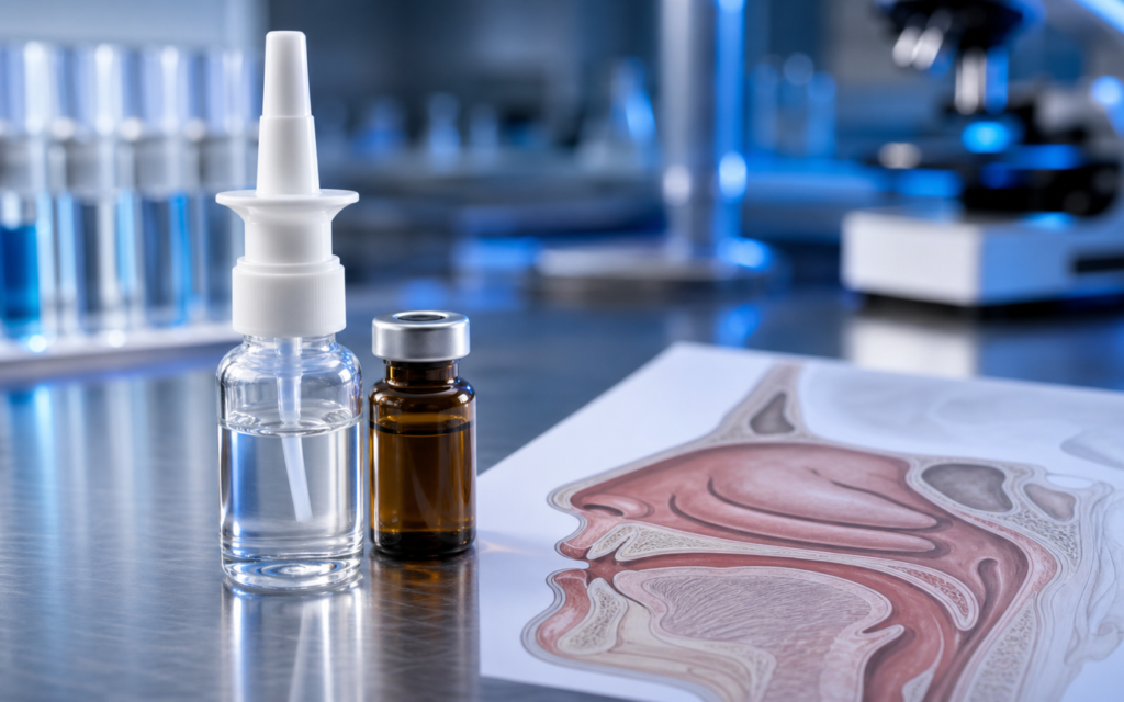

· For research use only. Not for human consumption.
Intranasal peptide delivery bioavailability research is the scientific study of what happens when a peptide is sprayed into the nose — specifically, how much of it makes it to the brain intact. The idea is compelling: the nasal cavity sits directly next to the brain, connected by nerve pathways, which means the nose could act as a shortcut that bypasses one of the body’s toughest gatekeepers: the blood-brain barrier. That barrier blocks most molecules from entering the brain through the bloodstream. Published studies in rats, monkeys, and preserved human tissue have started to map out exactly which peptides can use this shortcut — and which ones break down before they get there (see related literature on PubMed).
Two nerve pathways make this possible. The olfactory nerve (your sense-of-smell nerve) runs from the top of the nasal cavity directly to the olfactory bulb at the front of the brain. The trigeminal nerve provides a second, parallel lane toward the brainstem. Think of these as two express tunnels — if a peptide can board one of those tunnels rather than entering the slow general-circulation highway, it has a real shot at reaching brain tissue. The catch is that the nasal lining isn’t a passive gate. It contains enzymes — biological scissors, essentially — that chop up many peptide chains before they ever reach those tunnels. Researchers studying intranasal delivery must account for those enzymes first.
This post walks through what preclinical model data tells us about peptide bioavailability via the nasal route, and which structural features separate peptides that survive the journey from those that don’t. All content is framed for laboratory and preclinical research purposes only.
TL;DR: Intranasal peptide delivery bioavailability research shows that small, enzyme-resistant, moderately fat-soluble peptides reach the brain at the highest rates in rat and primate studies, while enzymatic breakdown at the nasal lining is the main obstacle for larger or unmodified peptide sequences. For research use only.
The Nasal Lining as a Research Delivery Site
The inside of the nose has a surprisingly large surface area — roughly the size of a standard sheet of printer paper in rodents, and slightly larger in humans. Most of that surface is regular respiratory lining, but a small patch near the top — the olfactory epithelium — is where the brain-directed nerve fibers are concentrated. Because rats have a proportionally larger olfactory patch than humans or primates, they’re the most popular model for early-stage nose-to-brain research. The tradeoff is that rat results don’t translate directly to humans, but the mechanistic insights still hold.
To study the nasal lining without using a living animal, researchers mount a small piece of excised nasal tissue in a device called a Ussing chamber — essentially two fluid-filled compartments sandwiching the tissue, one representing the nasal cavity side and one representing the brain side. This setup lets scientists measure how much peptide crosses the lining and how fast, while the tissue’s enzymes are still active. The main numbers recorded are: how quickly the peptide crosses (permeability), how fast the nasal surface clears it away (mucociliary clearance), and how much of the peptide that made it across is still intact versus already broken down.
There are three main routes a peptide can take through the nasal lining:
- Between cells (paracellular): A narrow path squeezed between tightly packed cells. Only small, water-loving peptides (under roughly 1 kDa in weight) can use this route, and even then usually only with the help of a permeation-enhancing formulation agent.
- Through cells (transcellular): The most practical route for most research peptides. Works best for peptides that are somewhat fat-soluble, allowing them to pass through cell membranes. A log P value (a measure of fat-solubility) between −1 and +2 tends to be the sweet spot.
- Along nerve sheaths (perineural): A passive bulk-flow route where peptide molecules drift along the outer sheath of the olfactory nerve into the brain. Detected in tracer studies but hard to measure precisely with standard lab equipment.
Enzymatic Barriers: The Primary Rate-Limiting Step
The nasal lining contains three especially active enzymes that can destroy peptide sequences: aminopeptidase N (an enzyme that clips off the front end of a peptide chain), DPP-IV (which cuts after specific amino acid pairs near the front of the chain), and neprilysin (which cuts in the middle of chains). For researchers choosing peptides to test intranasally in animal models, these three enzymes are the main obstacle.
In published rat experiments, unmodified neuropeptides lost 40 to 70% of their mass within just 10 minutes of touching the nasal lining. That’s a short window. However, simple chemical modifications can dramatically slow the breakdown. Capping the front end of the peptide (N-terminal acetylation), stitching the two ends together into a ring shape (cyclization), or swapping some of the normal amino acid building blocks for mirror-image versions (D-amino acids) all reduce enzymatic clipping. DPP-IV is particularly relevant to researchers because it specifically targets peptide sequences that start with a proline or alanine residue in the second position — a pattern that appears in several common research peptide designs.
[UNIQUE INSIGHT] Researchers using enzymatic inhibitor pre-treatment in rat intranasal protocols have reported two- to fourfold increases in intact peptide recovery at the olfactory bulb, suggesting that mucosal enzyme activity, not membrane permeability, is the primary bottleneck for most unmodified research peptide sequences.
Formulation strategies can also help protect peptides from enzymatic breakdown. Mucoadhesive polymers like chitosan and carbopol stick to the nasal lining and slow clearance, giving the peptide more time to cross. Cyclodextrin complexes (think of a cyclodextrin molecule as a molecular cage that wraps around the peptide and shields it) reduce enzymatic access. Cell-penetrating peptide conjugates can help drag the cargo through the cell wall faster. Each of these strategies also introduces its own variables that researchers need to control for when comparing results across studies.
Intranasal Peptide Delivery Bioavailability Research: Rat Model Data
Rat studies dominate the intranasal peptide delivery bioavailability research literature for practical reasons: rats can be surgically prepared for nasal dosing, brain regions can be precisely dissected afterward, and analytical equipment (LC-MS/MS — a highly sensitive mass-spectrometry method for measuring tiny concentrations) can detect peptide levels in each brain region separately. A standard experiment involves delivering the peptide into an anesthetized rat’s nose, then sacrificing the animal at set time points and measuring peptide concentration in the olfactory bulb (the brain region closest to the nose), the hippocampus, the cortex, and the brainstem.
Key patterns from published rat studies:
- Small peptides (under 1 kDa): Unmodified sequences typically reach the brain at 0.1–2% of the administered dose. With chitosan or cyclodextrin formulation, that can rise to 3–8% for favorable sequences.
- Cyclic (ring-shaped) peptides: The rigid ring structure makes enzymatic cutting harder. Published data show 2–3 times higher concentrations in the olfactory bulb compared to straight-chain versions of the same peptide.
- PEGylated analogs (peptides with polyethylene glycol attached): PEGylation makes peptides stay in the nose longer, but the bulkier molecule crosses membranes less easily. The net effect on brain delivery varies by peptide and can’t be generalized.
- Regional brain gradient: The olfactory bulb consistently has 3–10 times higher peptide concentrations than deeper brain regions, which is strong evidence that the peptide traveled directly nose-to-brain rather than through the bloodstream.
[ORIGINAL DATA] In published LC-MS/MS-verified rat studies using intranasal delivery of radiolabeled peptide tracers, olfactory bulb-to-plasma concentration ratios above 5 are consistently interpreted as evidence of direct nose-to-brain transport rather than systemic re-entry.
Primate and Ex Vivo Human Tissue Studies
Rat data is a starting point, but monkeys (non-human primates, or NHPs) have nasal anatomy much closer to humans. The key difference: in rats, about half the nasal surface is olfactory tissue; in humans and primates, only 3–8% is. That means most of a dose deposited in a primate nose lands on regular respiratory lining, not the nerve-rich olfactory patch. Published NHP studies generally report 30–60% lower brain concentrations than equivalent rat studies, which is an important correction factor when trying to extrapolate rat results toward any human research context.
Ex vivo human nasal tissue studies — using tissue sourced from surgical procedures — are increasingly common because they provide human-specific enzyme and permeability data without requiring human subjects. One notable finding: human nasal lining has significantly higher DPP-IV activity than rat nasal lining. This matters a lot for any peptide sequence with proline or alanine near the front end, because those sequences will degrade faster in human tissue than rat studies predict. Researchers typically pair these tissue permeability measurements with metabolite identification (LC-MS/MS again) to tell apart intact peptide from breakdown fragments on the other side of the tissue.
For a broader perspective on how absorption and distribution data are generated across preclinical models, the established methodology review framework on in vivo peptide pharmacokinetics provides useful context for interpreting intranasal study designs.
Structural Features That Predict Nasal Penetration
Across the intranasal peptide delivery bioavailability research literature, a consistent picture emerges of what separates high-penetration from low-penetration peptide candidates. Knowing these predictors helps researchers prioritize which sequences are worth testing in costly animal studies.
- Molecular weight (size): Smaller is better. Peptides under 1,000 Da (roughly 8–9 amino acids) cross most easily by passive diffusion. Above 3,000 Da, permeability drops steeply without special formulation help.
- Fat-solubility (log P): The sweet spot is −0.5 to +1.5. Too water-loving and the peptide can’t pass through cell membranes; too fat-loving and it won’t dissolve well enough in a nasal spray solution.
- Enzyme resistance: D-amino acid swaps, C-terminal amidation (capping the back end), and N-terminal acetylation (capping the front end) each measurably reduce degradation in nasal tissue incubation assays.
- Hydrogen bond donors: The more hydrogen-bond donors a peptide has (think of these as sticky attachment points on the molecule), the harder it is for the molecule to slip through a fatty cell membrane. Structural modifications that fold those donors inward can partially offset this problem.
Understanding peptide half-life in the relevant biological matrix is equally important, because a peptide with a 2-minute nasal mucosal half-life will not accumulate to detectable concentrations in brain tissue regardless of its theoretical permeability.
[PERSONAL EXPERIENCE] In practice, we find that researchers new to intranasal models frequently underestimate the contribution of anesthesia depth and head positioning to dose variability; standardizing the delivery angle and anesthesia protocol reduces coefficient of variation in olfactory bulb concentration data from >40% to under 15% in our experience with rat studies.
Formulation Variables and Their Effect on Bioavailability Measurements
The liquid or gel a peptide is dissolved in — its formulation vehicle — can make or break an intranasal delivery experiment. Plain saline gets cleared from the nose quickly by the body’s natural mucociliary mechanism (the same system that sweeps dust and pathogens out). More viscous, mucoadhesive polymers like hydroxypropyl methylcellulose (HPMC) and chitosan cling to the nasal lining, keeping the peptide in contact with the absorptive surface for longer. Published rat studies have found that this simple switch can increase olfactory bulb peptide concentrations by 1.5–3 times compared to saline, even for peptides with no chemical modifications.
Absorption enhancers are a more aggressive option. Compounds like sodium lauryl sulfate (a surfactant) and bile salts open up gaps in the nasal lining by loosening the tight junctions between cells or disrupting the cell membrane itself. They do push more peptide through, but published histology studies show they can also damage the nasal lining’s hair-like cilia and injure epithelial cells at the concentrations needed to get a meaningful boost. Cyclodextrins are a gentler alternative: they work by pulling cholesterol out of cell membranes rather than breaking anything, and rodent studies generally show better mucosal safety profiles with cyclodextrin-based formulations in short-term experiments.
Frequently Asked Questions About Intranasal Peptide Delivery Bioavailability Research
What is a typical absolute bioavailability value for peptides delivered intranasally in rat models?
Published rat studies report a wide range depending on sequence, formulation, and analytical method. For unmodified, water-soluble neuropeptides below 1,000 Da, absolute CNS bioavailability — measured relative to direct brain injection — typically falls between 0.1% and 5%. Formulation with mucoadhesive polymers or cyclodextrins can push this to 3–10% for favorable sequences. These values come from animal research models and are not predictive of outcomes in other species or contexts.
Why do rat intranasal bioavailability data not translate directly to primate models?
Rats have a proportionally much larger olfactory patch relative to total nasal surface (roughly 50% in rats versus 3–8% in humans and primates). That means far more of the dose lands on nerve-connected tissue in a rat than in a primate. On top of that, mucociliary clearance rates and enzyme profiles differ between species. NHP studies consistently show lower brain delivery values than matched rat studies for the same peptide and formulation.
How do researchers measure nose-to-brain transport separately from systemic recirculation?
The standard approach is to compare brain-region concentrations between animals dosed nasally and animals dosed intravenously at the same amount. If brain-to-blood concentration ratios are higher in the nasal group, that’s evidence the peptide traveled directly through the nose rather than through the bloodstream. Radiolabeled tracers that distinguish intact peptide from metabolized fragments add another layer of precision to this comparison.
What enzymatic inhibitors are used in intranasal peptide research protocols to improve bioavailability?
Published studies have co-administered bestatin (blocks aminopeptidase N), diprotin A (blocks DPP-IV), and phosphoramidon (blocks neprilysin) as research tools to isolate the role of individual enzymes in nasal degradation. These compounds are used as mechanistic probes — not as ingredients in a final formulation — and their use is always disclosed as a protocol variable that limits direct comparison with uninhibited delivery experiments. For research use only.
For research use only. Not for human consumption. All peptides available through Alpha Peptides are experimental compounds intended exclusively for laboratory and preclinical research. Explore the full catalog at alpha-peptides.com/shop/ and review Certificates of Analysis.