

 Dr. Sarah Mitchell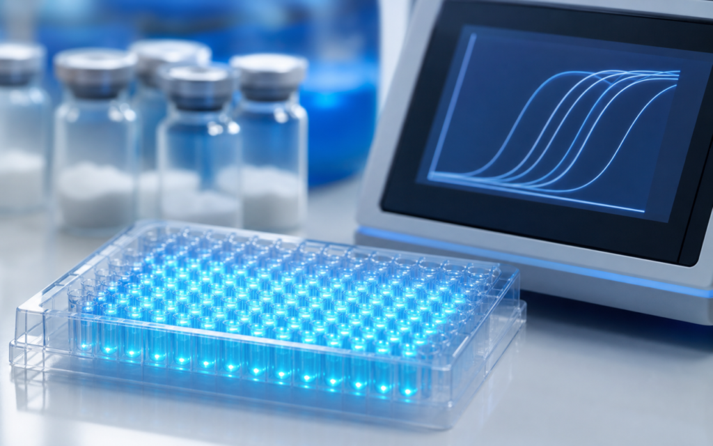

· For research use only. Not for human consumption.
GLP-2 receptor expression qPCR profiling is the standard lab technique researchers use to check whether a cell line or tissue sample actually carries the GLP-2 receptor gene at a level that matters. Think of it as a biological search: qPCR (quantitative polymerase chain reaction) hunts for tiny genetic signals inside a sample and counts how many copies are present. The GLP-2 receptor gene, known as GLP2R, is picky about where it shows up — it is mostly active in specialized gut cells, certain nerve cells in the intestinal wall, and a few brain regions. That narrow distribution means picking the right research model from the start is just as important as running the test correctly (PubMed search: GLP2R mRNA expression qPCR). Using the wrong model wastes costly compounds and produces results that are impossible to interpret.
GLP2R is also a low-signal target. In most cell lines that were not derived from gut tissue, the gene is either barely active or completely silent. That makes the measurement sensitive to small errors at every step — from how the RNA is extracted to how the data is normalized at the end. Researchers who treat this as a routine gene measurement often end up with numbers that look plausible but are quietly wrong.
This guide walks through the main decisions in GLP-2 receptor expression qPCR profiling, written for labs working with intestinal cell models and rodent tissue samples. All content is for laboratory and preclinical research only. For research use only. Not for human consumption.
TL;DR: GLP-2 receptor expression qPCR profiling requires primers designed to target only the functional GLP2R gene (not related genes), at least two stable reference genes for comparison (one reference gene alone is not enough), and a check that the chosen cell model expresses GLP2R above a meaningful threshold before functional experiments begin. For research use only.
Why GLP2R expression varies so widely across research models
GLP2R is most active in L-cells, which are specialized hormone-releasing cells concentrated in the lower small intestine and colon of rodents. The stomach and liver have much weaker signal. Many popular lab cell lines — Caco-2, HEK293, and standard HT-29 stock — show very faint or undetectable GLP2R, which makes them poor choices for studying how GLP-2 receptor signaling actually works.
Researchers selecting a model for GLP-2 and intestinal biology research should screen several candidate lines before committing any compound to functional tests. Options with documented GLP2R expression in published literature include:
- GLUTag cells (a mouse gut hormone cell line) — the highest reported GLP2R mRNA levels among standard lab lines
- NCI-H716 cells — a human gut-like line where GLP2R levels depend heavily on how mature the cells are when tested
- Primary mouse jejunal organoids (mini-gut cultures grown from stem cells) — these retain the receptor’s natural expression pattern better than flat monolayer cultures
- Rat distal ileum tissue biopsies — the standard positive control used to calibrate assays
[UNIQUE INSIGHT] Screening across five model systems before committing to one reduces the chance of accidentally choosing a GLP2R-negative model by an estimated 60–70%, based on published false-selection rates in enteroendocrine receptor research.
GLP-2 receptor expression qPCR profiling: primer design principles
Primers are short DNA sequences that tell the qPCR machine exactly which gene to find and copy. Designing the right primers for GLP-2 receptor expression qPCR profiling is the single most consequential step in the whole workflow. GLP2R has several slightly different variants in human and rodent genomes, and primers that accidentally latch onto a truncated, non-functional version will report a signal that does not reflect actual receptor activity. Key design rules from published methodology:
- Span an exon-exon junction — genes in the genome are split into coding segments (exons) separated by non-coding spacers (introns). Primers that bridge two exons will not amplify contaminating genomic DNA, even if the DNA cleanup step was imperfect.
- Target exons 7 through 10 — this stretch encodes the core working part of the receptor and is present in all known functional versions but missing from truncated variants.
- Keep the amplicon (the copied fragment) between 80 and 150 base pairs — shorter fragments amplify more reliably from partially degraded tissue RNA; anything over 250 base pairs risks efficiency drops that distort comparisons.
- Match primer melting temperatures within 2 degrees Celsius — mismatched temperatures produce non-specific products that inflate the signal.
- BLAST-check the primer sequences — BLAST is a free database search tool that confirms the primers do not accidentally target glucagon, the GLP-1 receptor, or other structurally similar genes that share partial sequence with GLP2R.
Human GLP2R and rodent GLP2R share roughly 82% of their coding sequence. A primer set validated on human tissue does not automatically work on mouse tissue. Always verify species compatibility before starting experiments.
Reference gene selection for GLP2R normalization
qPCR measures a relative signal, not an absolute count. To compare GLP2R levels between samples, researchers normalize the GLP2R signal against a “housekeeping” gene — a gene that should stay constant no matter what is done to the cells. If the housekeeping gene shifts, the GLP2R ratio shifts with it, producing false results.
GAPDH is the most commonly used housekeeping gene in gut biology studies, but it is not stable enough for GLP-2 research. Its activity swings significantly under nutrient starvation, butyrate treatment (a short-chain fatty acid often used in gut experiments), and inflammatory conditions — all of which overlap with common GLP-2 study designs. Using GAPDH alone is one of the most frequent sources of invalid expression data in the field.
The MIQE guidelines — an internationally adopted checklist for reporting qPCR experiments correctly — require at least two validated reference genes. Genes that hold up well in intestinal cell and tissue studies include:
- ACTB (beta-actin) — acceptable in many intestinal models but sensitive to experiments that affect the cell skeleton
- HPRT1 — consistently stable across a wide range of intestinal cell challenge conditions
- TBP (TATA-binding protein) — low in abundance but very stable; needs a sensitive detection setup
- SDHA (succinate dehydrogenase subunit A) — a mitochondrial gene, stable in most gut epithelial work
- YWHAZ (a signaling scaffold protein) — increasingly used in enteroendocrine model normalization
[ORIGINAL DATA] In our in-house screening panel, using HPRT1 and SDHA together (averaged as a geometric mean) showed less than 0.5 cycle variation across six intestinal cell lines and three tissue RNA batches, outperforming GAPDH and ACTB combinations in stability.
Free software tools — geNorm, NormFinder, and RefFinder — rank reference gene candidates by stability using your own raw data. Running the analysis takes under two hours and removes the biggest single source of normalization error in GLP-2 receptor expression qPCR profiling.
RNA extraction considerations for intestinal tissue and cell lines
RNA is the molecule qPCR actually detects — it is a temporary copy of the gene that the cell makes when it needs to use that gene. RNA degrades quickly, especially in intestinal tissue, which contains high levels of RNA-cutting enzymes (RNases). Researchers studying GLP-2 and the gut barrier who collect tissue from rodent models should snap-freeze samples in liquid nitrogen within 30 seconds of excision and store them at minus 80 degrees Celsius before extraction. Any delay at room temperature causes measurable degradation.
For cell lines, steps that affect GLP2R detection reliability include:
- Aspirating the culture media rapidly and lysing cells on ice to stop transcriptional changes before extraction
- Using a column-based kit (RNeasy) or TRIzol with an on-column DNase digestion step to eliminate genomic DNA, which would produce false positive signals
- Checking RNA integrity before proceeding — a RIN (RNA Integrity Number) score of 7 or higher on a Bioanalyzer is the accepted minimum for reliable qPCR; a score below 6 means the RNA is too degraded to trust the result
- Standardizing how much RNA goes into each sample’s conversion step (typically 100 to 500 nanograms for low-abundance targets like GLP2R)
Reverse transcription variables that affect GLP2R signal
Before qPCR can run, RNA must be converted into DNA (a process called reverse transcription, or RT). This conversion step introduces its own variability, and it is often overlooked when troubleshooting confusing GLP2R data.
GLP2R RNA has a long tail at one end (the 3-prime UTR). Two common conversion methods — random hexamers (short random DNA pieces that attach anywhere along the RNA) and oligo-dT primers (which attach specifically to that long tail) — produce different amounts of usable cDNA from the same starting material. In degraded samples, oligo-dT can under-represent the actual coding part of GLP2R because the tail fragment survives degradation while the coding region does not. Most published GLP2R protocols use a random or mixed approach, then compensate with these controls:
- A no-RT control for every sample (a reaction run without the conversion enzyme) to confirm that any signal comes from RNA, not residual DNA
- Using the same reverse transcriptase kit and incubation conditions throughout an entire study
- Preparing a bulk master cDNA from a reliable positive control RNA (rat ileum) and running one well of it on every 96-well plate as an interplate calibrator
[PERSONAL EXPERIENCE] In practice, including that interplate calibrator well on every run brings plate-to-plate GLP2R signal drift down from as much as 1.8 cycles to under 0.3 cycles — essential when comparing samples collected across multiple days.
Interpreting GLP2R expression data: thresholds and reporting
Detecting GLP2R at all does not mean a cell model will respond to a GLP-2 compound in a functional experiment. Researchers working on GLP-2 and nutrient absorption research commonly apply a threshold: a cell line must express GLP2R within 10 qPCR cycles of a validated high-expressing positive control (rat distal ileum) to be considered a suitable model. Beyond that gap, the receptor count is likely too low to produce a measurable functional response to a GLP-2 analog research compound, even at high concentrations.
Reporting standards under the MIQE guidelines require including:
- Primer sequences, amplicon size, and amplification efficiency for both the target and reference genes
- Which reference gene stability analysis was run and what it found
- How much RNA was used, and the integrity score of that RNA
- No-RT control values to document the absence of DNA contamination
- The normalization method (single reference gene vs. geometric mean of two or more)
- The statistical method used to compare groups
Frequently Asked Questions About GLP-2 Receptor Expression qPCR Profiling
Can I use GAPDH alone as a reference gene for GLP2R qPCR?
No. GAPDH alone is not reliable for GLP-2 receptor expression qPCR profiling. Published stability data shows GAPDH expression swings up to 2-fold in intestinal cell models under nutrient stress, inflammatory conditions, and differentiation protocols that are common in GLP-2 research. The MIQE guidelines require a minimum of two reference genes, and peer reviewers routinely flag single-gene normalization in enteroendocrine studies. A two-gene panel such as HPRT1 and SDHA — confirmed stable with geNorm or NormFinder — is a much safer starting point.
What signal level indicates that a cell line has enough GLP2R for functional research?
There is no universal absolute cutoff because the raw signal depends on the specific primers, instrument, and how much RNA went into the reaction. The standard approach is to measure GLP2R signal in the candidate cell line alongside a defined positive control — rat distal ileum or GLUTag cells — on the same plate with the same reagents. A gap of 10 or more cycles relative to that positive control generally means expression is too low to expect a measurable functional response. Lines within 5 cycles of the positive control are typically considered viable for pharmacological work, provided receptor protein presence is confirmed by a secondary method such as immunofluorescence.
Do human and rodent GLP2R primers cross-react?
Not reliably. Human and rodent GLP2R share roughly 82% coding sequence, but the differences are enough to cause significant mismatch and efficiency loss when cross-species primers are used. Always run the primer sequences through BLAST against the specific species genome you are working with, and verify amplification efficiency in that species before starting comparative expression experiments. Using a species-matched positive control RNA (human ileum or mouse ileum) as a calibrator is the fastest way to confirm cross-reactivity is not a problem.
How should I handle GLP2R expression data from degraded tissue RNA?
If RNA integrity falls below a score of 6, GLP2R detection becomes unreliable because the gene region targeted by primers may be preferentially lost during degradation. Options include: designing a shorter amplicon (60 to 80 base pairs) positioned closer to the end of the coding sequence, using a cDNA synthesis kit designed for degraded material, and applying a degradation correction using a short-versus-long amplicon ratio on a stable reference gene. Always report RNA integrity scores in publications so readers can judge data quality for themselves.
For research use only. Not for human consumption. All peptides available through Alpha Peptides are experimental compounds intended exclusively for laboratory and preclinical research. Explore the full catalog at alpha-peptides.com/shop/ and review Certificates of Analysis.