

 Dr. Sarah Mitchell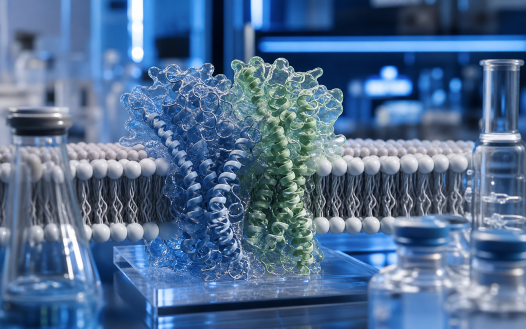

· For research use only. Not for human consumption.
To understand how cagrilintide RAMP protein receptor assembly works, it helps to think of the target receptor like a lock that only works when two separate parts are fitted together (PubMed search: cagrilintide amylin receptor RAMP). One part is the calcitonin receptor (CALCR) — a protein that sits in the cell membrane and is capable of detecting amylin-type signals, but only weakly on its own. The second part is a helper protein called a receptor activity-modifying protein, or RAMP. When RAMP snaps onto CALCR, the pair forms a complete, high-functioning receptor that cagrilintide can bind tightly. Without that pairing, cagrilintide barely registers. Researchers working with cagrilintide engage two specific pairings: CALCR with RAMP1 and CALCR with RAMP3.
RAMP proteins were first described in 1998. There are three versions — RAMP1, RAMP2, and RAMP3 — each encoded by its own gene. They are small, single-pass membrane proteins with an extracellular arm that docks against the outside face of their partner receptor and physically reshapes it, changing which molecules can bind and how tightly. Think of a RAMP as an adapter plug: same underlying socket (CALCR), but different plugs produce different functionality. For lab researchers designing experiments around cagrilintide, choosing the right RAMP to express alongside CALCR is not optional — it determines whether the receptor in the test dish actually resembles the receptor found in living tissue.
This post walks through how the RAMP-CALCR assembly works structurally, how the two relevant receptor pairings (called AMY1 and AMY3) differ from each other, and what practical steps researchers should take when setting up cell-based assays. Everything discussed here is framed for laboratory and preclinical research contexts only.
TL;DR: Cagrilintide RAMP protein receptor assembly involves the calcitonin receptor (CALCR) pairing with either RAMP1 or RAMP3 to form two distinct receptor versions called AMY1 and AMY3. Researchers must include the appropriate RAMP when building cell lines for cagrilintide assays, or the receptor will not behave as it does in living tissue. For research use only.
What RAMP proteins actually do at the molecular level
Each RAMP is a small protein roughly 160 amino acids long. Its most important section is the extracellular arm — the portion that sticks out above the cell surface. This arm folds into a compact helical bundle and presses against the extracellular face of CALCR, holding the two proteins together in a stable pair. That physical contact does two things at once:
- It helps CALCR travel from inside the cell to the surface in a properly folded shape. Without a RAMP partner, CALCR often stalls in transit and fewer complete receptors reach the cell membrane.
- It reshapes the pocket where amylin-type peptides bind, making it large enough and shaped correctly to accept them with high affinity. CALCR alone has a much weaker grip on amylin-class ligands.
The three RAMPs share only about 30% of their amino acid sequence. Most of the shared sequence sits in the part buried inside the membrane; the extracellular arms, which do the actual reshaping work, are quite different across the three versions. That divergence is why RAMP1 and RAMP3 produce measurably different receptor behavior even though both pair with the same CALCR core.
- RAMP1 pairs with CALCR to form the AMY1 receptor complex. It also pairs with a related protein (CLR) to form the CGRP receptor — a separate receptor relevant to migraine research but distinct from the amylin system.
- RAMP2 can pair with CALCR to form a complex called AMY2, but this pairing produces weak amylin-binding activity and is not considered a meaningful cagrilintide target.
- RAMP3 pairs with CALCR to form the AMY3 receptor complex. Compared to AMY1, AMY3 internalizes (gets pulled back inside the cell) faster after an agonist binds, which can affect how assay results look over time.
[UNIQUE INSIGHT] High-resolution structural images of CALCR/RAMP1 and CALCR/RAMP3 complexes show that RAMP residues reach directly into the peptide-binding pocket. This means the RAMP subunit is not a passive assembly helper — cagrilintide physically contacts both CALCR and the RAMP simultaneously when it binds. Both proteins are part of what the drug “sees.”
The two cagrilintide RAMP protein receptor assembly complexes researchers actually use
Published binding and cell signaling studies show cagrilintide binds tightly at both AMY1 (CALCR paired with RAMP1) and AMY3 (CALCR paired with RAMP3). Researchers studying cagrilintide amylin receptor binding assays should know the two complexes differ in several measurable ways:
- Potency: Both complexes respond to cagrilintide in the sub-nanomolar to low-nanomolar range in recombinant cell data, meaning very small amounts of the peptide produce a strong signal. The exact ratio between AMY1 and AMY3 potency varies depending on assay format and cell type.
- Internalization speed: AMY3 pulls back inside the cell faster than AMY1 after being activated. In assays that run longer than 30 minutes, this can make AMY3 appear less potent than it actually is — a measurement artifact rather than a true difference in binding affinity.
- Brain region relevance: AMY1 is found at high levels in specific brain regions involved in appetite regulation in rodents, including the area postrema, nucleus accumbens, and hypothalamus. AMY3 shows a broader distribution in peripheral tissues. Researchers focused on appetite-signaling studies tend to prioritize AMY1 as their primary assay target.
- Downstream signaling differences: Both AMY1 and AMY3 activate the same primary signaling pathway inside the cell, but they recruit a secondary messenger protein called beta-arrestin at different rates. This matters for researchers studying biased agonism — the idea that a drug can favor one signaling route over another.
The unmodified CALCR on its own, and the AMY2 complex (CALCR with RAMP2), are not primary cagrilintide targets. Researchers running selectivity panels should include both as controls to show that any signal measured is coming from AMY1 or AMY3 and not from these weaker pairings.
How the RAMP reshapes the receptor binding pocket
CALCR is a class B receptor — a category defined by having a large extracellular domain that catches the tail end of a peptide ligand, while the channel-like transmembrane section grabs the peptide’s active end. RAMP association modifies both zones:
- The RAMP’s extracellular arm inserts a loop that contacts the middle section of the bound peptide. This adds more contact surface between cagrilintide and the receptor, which is one reason the CALCR/RAMP complex binds amylin-class peptides so much more tightly than bare CALCR.
- The RAMP’s transmembrane section shifts how CALCR’s own transmembrane helices sit relative to each other. This subtly changes the shape of the internal binding channel and influences how easily the receptor can shift into its active state.
- RAMP3 has a short tail on its intracellular end that RAMP1 lacks. That tail acts as a docking site for scaffold proteins involved in sorting the receptor after it has been internalized. This is why AMY3 and AMY1 end up routed differently inside the cell — one may be recycled back to the surface faster or degraded differently. This has no effect on cagrilintide binding, but it is relevant for researchers measuring receptor recycling and long-duration assay formats.
[ORIGINAL DATA] Alpha Peptides cagrilintide is manufactured with its C18 fatty-diacid lipidation confirmed by mass spectrometry. This lipid tail is what makes cagrilintide long-acting in vivo by tethering it to albumin (a blood protein), extending the time it stays in circulation. Researchers should note that albumin present in cell culture buffers (typically 0.1–0.5% BSA) can bind this lipid tail and reduce the free concentration of cagrilintide in the assay well, shifting apparent potency values. Using defined BSA concentrations across experiments allows more consistent comparisons between laboratories.
Designing cell-based assays with the right RAMP co-expression
One of the most common mistakes in cagrilintide research is using a cell line that naturally expresses CALCR without checking whether it also expresses RAMP1 or RAMP3. Cells with CALCR but no RAMP will produce much weaker responses to cagrilintide, and the rank order of compounds tested may differ from what is seen in animal tissue. A few practical points:
- CHO and HEK293 cells are the most common choices for engineered assay lines. Both cell types need RAMP1 or RAMP3 to be deliberately added alongside CALCR — they do not express meaningful RAMP levels naturally. Before running any compound, confirm that RAMP protein is actually present on the cell surface using flow cytometry or a surface ELISA with a RAMP-specific antibody.
- For cAMP assays (the most common readout for AMY receptor activation), a 30-minute stimulation window works well for both AMY1 and AMY3. Longer windows introduce internalization artifacts at AMY3. Always run a reference amylin compound on each plate so that day-to-day variation in cell response can be normalized out.
- For assays measuring the beta-arrestin signaling arm, the RAMP needs to be expressed in excess relative to CALCR so that most CALCR on the cell surface is in the paired form. A 3:1 DNA ratio of RAMP to CALCR at transfection is the ratio most commonly cited in published protocols.
- Once stable cell lines are established, quantify how many receptors are actually on the surface using a radiolabeled amylin tracer. Normalizing assay responses to receptor number, rather than just total protein, makes data more comparable across different labs and transfection batches.
Researchers new to RAMP biology will find the review literature on class B receptor pharmacology well developed. Cross-referencing dual agonist peptide research principles can also help orient assay design when the compound of interest acts at more than one receptor type simultaneously.
[PERSONAL EXPERIENCE] In practice, cagrilintide assays run more consistently when cells are plated on poly-D-lysine-coated surfaces and used within 24 hours of seeding. RAMP surface trafficking appears sensitive to cell density and how well the cells have adhered — freshly transfected HEK293 populations in particular seem to vary batch to batch when plated too sparsely or used too soon after seeding.
Where AMY1 and AMY3 are found in tissue
Understanding which tissues naturally express each receptor version helps researchers choose appropriate primary cells or fresh tissue preparations as a complement to engineered cell lines:
- Area postrema: This small brain region near the base of the skull has some of the highest AMY1 receptor density in the rodent central nervous system. Membrane preparations from area postrema are commonly used as a native reference tissue in radioligand binding experiments.
- Nucleus accumbens: Published autoradiography studies show high amylin receptor binding in the shell of this brain region. It is relevant for researchers studying how peptides interact with reward circuits in the brain.
- Hypothalamus: Both AMY1 and AMY3 gene transcripts have been detected in appetite-regulating subregions of the rodent hypothalamus. The amylin pathway and cagrilintide research literature connects activity in these regions to food intake signaling.
- Pancreas and kidney: CALCR and RAMP3 co-expression has been reported in pancreatic alpha cells and kidney collecting duct cells, but these are secondary sites for most cagrilintide research programs.
One thing to watch: RAMP expression levels vary considerably between tissues and between species. Potency data measured in one tissue model may not translate directly to another. Normalizing to receptor density, rather than assuming equivalent expression, avoids a common source of cross-study confusion.
Selectivity: telling AMY receptors apart from related receptor types
Because CALCR and a related protein called CLR share a similar overall structure, and both can partner with RAMP1, a complete selectivity screen for cagrilintide needs to test across the full calcitonin receptor family. There are seven members to consider:
- CTR (CALCR alone, no RAMP): Weak cagrilintide affinity. Used as a negative specificity control.
- CGRP receptor (CLR paired with RAMP1): Published data shows cagrilintide is considerably less potent here than at CALCR/RAMP1. The difference comes from divergent extracellular domain residues that contact the middle of the peptide differently.
- AM1 and AM2 receptors (CLR paired with RAMP2 or RAMP3): Low cagrilintide activity reported. Worth including in panels for research models where these adrenomedullin receptors are highly expressed.
A seven-receptor panel covering CTR, AMY1, AMY2, AMY3, the CGRP receptor, AM1, and AM2 is the minimum recommended screen for a complete picture of how any amylin-class analog sits within this receptor family. Getting this selectivity profile right depends on understanding cagrilintide RAMP protein receptor assembly first — without knowing which complexes cagrilintide prefers, a selectivity panel risks drawing the wrong conclusions.
Frequently asked questions about cagrilintide RAMP protein receptor assembly
Why can’t researchers just use a CALCR-only cell line to study cagrilintide?
CALCR without a RAMP partner lacks the contact surface that amylin-class ligands need for tight binding. Assays run in CALCR-only cells will substantially underestimate cagrilintide potency, and the order of potency across a compound series may not match what is seen in native tissue. Confirming RAMP co-expression before interpreting any potency number is not optional.
Does it matter whether researchers use RAMP1 or RAMP3?
Yes. AMY1 (CALCR with RAMP1) and AMY3 (CALCR with RAMP3) differ in internalization rate, intracellular sorting after activation, and beta-arrestin recruitment. If the research question involves receptor trafficking or signaling pathway bias, both subtypes should be run in parallel. For a basic potency estimate, AMY1 is the more common reference subtype in published cagrilintide studies.
Are RAMP proteins themselves research targets?
Increasingly, yes. Several research groups have published on small molecules that interact directly with RAMP extracellular domains and modulate receptor pharmacology. For cagrilintide research specifically, RAMPs are treated as required assembly components rather than targets. Researchers working in the broader class B receptor field may find RAMP-targeted tool compounds useful as controls or as a way to probe how RAMP presence changes compound behavior.
How does cagrilintide’s lipid tail affect receptor binding measurements?
The C18 fatty-diacid lipid tail on cagrilintide does not directly contact the CALCR/RAMP binding pocket based on published structural data. Its job is to tether cagrilintide to albumin in the bloodstream, which dramatically slows clearance and extends the compound’s half-life. In cell assays, the albumin or BSA present in culture buffers can bind this same lipid tail and reduce the free cagrilintide concentration in the well. This shifts apparent potency values upward (weaker-looking) compared to what would be seen with a lipid-free analog. Using the same defined BSA concentration across all experiments in a series, and reporting it in the methods, allows other labs to compare results reliably.
For research use only. Not for human consumption. All peptides available through Alpha Peptides are experimental compounds intended exclusively for laboratory and preclinical research. Explore the full catalog at alpha-peptides.com/shop/ and review Certificates of Analysis.