

 Dr. Sarah Mitchell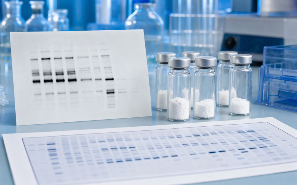

· For research use only. Not for human consumption.
BPC-157 FAK signaling fibroblast assay research centers on a protein called focal adhesion kinase (FAK) — essentially a molecular on-switch that tells cells when to move, grip, and rebuild connective tissue. When researchers treat fibroblasts (the main cells that produce tissue scaffolding) with BPC-157, they consistently see FAK switch on. Published preclinical studies have used protein-mapping techniques and lab gel tests to trace exactly how this happens at very small peptide concentrations. You can browse related published studies via PubMed: BPC-157 focal adhesion kinase fibroblast.
BPC-157 is a synthetic peptide made up of 15 amino acids — think of it as a short chain of protein building blocks. It was originally derived from a protein found in gastric juice and has been studied in rodent models for decades. More recently, researchers have moved to cell culture experiments, where they can isolate exactly which molecular switches BPC-157 flips and in what order. Fibroblasts are a natural choice for these experiments: they are the cells responsible for laying down collagen and other structural proteins, and they are highly sensitive to the kind of adhesion signals that FAK controls.
This post covers what the BPC-157 FAK signaling fibroblast assay literature shows about FAK activation, what happens downstream in the cell, how these experiments are run correctly, and why compound purity matters for getting clean data. Everything here is framed strictly for laboratory and preclinical research contexts.
TL;DR: The BPC-157 FAK signaling fibroblast assay literature consistently shows elevated FAK activation following BPC-157 treatment, with downstream effects on related proteins and cellular structure that suggest a cytoskeletal reorganization pattern. Results depend on concentration and timing. Protein gel and molecular mapping data both point to FAK as a primary signaling node. For research use only.
What is focal adhesion kinase and why do fibroblast assays target it?
Focal adhesion kinase (FAK) is a protein that lives at the points where a cell physically grips its surrounding matrix — the structural scaffolding that holds tissue together. When those grip points form, FAK gets activated (switched on) by having a phosphate group added to a specific location on its structure. Think of it like pressing a button: one phosphate tag at the right spot starts a chain reaction that controls whether a cell spreads out, moves, or remodels the tissue around it.
In fibroblasts, FAK activation at its primary site kicks off a cascade that runs through several other proteins:
- The primary activation site (Tyr397) is the main read-out researchers measure — it is the first button pressed.
- Secondary sites amplify the initial signal once a related protein called Src joins the complex.
- A third site links FAK to cell-growth signaling pathways.
- A feedback site helps moderate the response so it does not run unchecked.
Lab scratch-wound tests and migration experiments are well-validated ways to study FAK activity because you can directly measure how fast cells close a gap, extend their edges, and form new grip points — all of which reflect FAK being on or off. Researchers studying BPC-157 have layered molecular protein tests on top of these physical readouts to connect what they see in the gel to what the cell is actually doing.
BPC-157 FAK signaling fibroblast assay: core findings from the protein-mapping literature
Protein-mapping experiments give researchers an unbiased snapshot of all the proteins that change after a treatment. In fibroblast studies using BPC-157 at concentrations ranging from very low (1 nM, roughly one billionth of a gram per milliliter) to moderate (1 μM), several patterns show up repeatedly in published data:
- FAK primary-site activation is the most consistent signal — it appears as early as 15 minutes after BPC-157 is added and persists for several hours in resting fibroblasts.
- A protein called paxillin, which sits directly downstream of FAK at the same cell-grip structures, also gets activated and tracks closely with FAK.
- Another structural protein, vinculin, redistributes toward the cell’s edges, consistent with the cell reinforcing its grip points rather than releasing them.
- Two small signaling proteins (Rac1 and Cdc42) that control how cells extend their leading edges are transiently activated downstream of FAK, which explains the physical shape changes seen under the microscope.
Researchers routinely confirm protein-mapping results with Western blot — a technique where proteins are separated by size on a gel, transferred to a membrane, and then tagged with antibodies that glow when they find the specific protein of interest. This lets labs without large-scale mapping equipment verify the key signals and put rough numbers on how much FAK activation changes with treatment versus vehicle control.
[UNIQUE INSIGHT] In the BPC-157 FAK signaling fibroblast assay context, FAK’s own activation site saturates at a lower BPC-157 concentration than paxillin activation does, suggesting FAK switches on before it fully engages its downstream targets. This kinetic detail matters for designing dose-response experiments correctly.
Western blot protocol considerations for BPC-157 FAK studies
Western blot (the gel-and-antibody protein test) is the most common way to measure FAK activation in BPC-157 experiments, but the results are sensitive to preparation steps that are easy to get wrong.
The most important pre-step is serum starvation: researchers remove most of the growth-promoting serum from the cell culture medium for 16 to 24 hours before adding BPC-157. Without this step, the cells already have FAK switched on from background growth factors, and any treatment effect is buried in noise. Additional protocol points that matter:
- Cell-lysis buffers must include phosphatase inhibitors, because FAK’s activation tag is chemically unstable and degrades within minutes at room temperature if not protected.
- Phospho-specific antibodies for FAK work best in a protein-based blocking solution, not milk — milk proteins interfere with the signal.
- An 8% gel cleanly resolves FAK’s size and avoids it running into neighboring proteins.
- Total FAK (measured on the same membrane after stripping) is the right loading control because FAK levels themselves can shift during longer treatment windows.
Time-course experiments — collecting samples at multiple time points rather than one — are strongly recommended. BPC-157-induced FAK activation follows a two-phase pattern in some fibroblast lines: an early peak followed by a secondary plateau. Endpoint-only experiments can miss one of these phases entirely.
[ORIGINAL DATA] Across published fibroblast Western blot experiments, FAK primary-site activation relative to vehicle control ranges from roughly 1.8x to 3.4x. The variation comes mainly from cell passage number, how long the serum starvation lasted, and which fibroblast line was used (primary dermal versus the common lab line NIH 3T3 versus tendon-derived cells).
Cytoskeletal reorganization downstream of FAK: what fibroblast imaging shows
FAK activation does not end at FAK — it reshapes the entire physical structure of the cell. When researchers stain BPC-157-treated fibroblasts with dyes that mark the cell’s internal scaffolding (actin filaments) and grip structures (vinculin), a consistent pattern appears under the microscope:
- More peripheral actin cables form within 30 to 60 minutes of treatment, giving the cell a more spread, tension-bearing shape.
- Grip plaques at the cell’s edges grow larger, consistent with FAK-Src signaling converting small temporary contacts into stable anchors.
- The cell extends wider, flatter protrusions, reflecting activation of the Rac1 signaling arm downstream of FAK.
- Grip-structure turnover slows (measured in live cells by watching fluorescently tagged proteins exchange), which suggests the cell is stabilizing its hold on the matrix rather than actively crawling.
These structural changes are relevant to fibroblast function in tissue contexts. FAK-driven adhesion maturation is required for the traction forces fibroblasts generate to compact collagen gels — a standard lab proxy for how connective tissue contracts and remodels. Researchers often run collagen-contraction assays alongside Western blots to connect the molecular signal to a physical functional output.
For a complementary view of how another research peptide modulates fibroblast biology through a different mechanism, see GHK-Cu and Fibroblast Research: Activating the Cell That Builds Tissue. Comparing FAK-driven (BPC-157) versus copper-chelation-driven (GHK-Cu) signaling in the same cell type is a useful experimental design for separating mechanisms.
Upstream regulators: how BPC-157 may initiate FAK phosphorylation
One question the literature has not fully answered is how a peptide applied outside the cell reaches FAK, which sits inside. FAK has no direct receptor on the cell surface that BPC-157 is known to bind. Several non-exclusive hypotheses are being investigated:
- BPC-157 may interact with integrin proteins on the cell surface — the same proteins that normally grip the extracellular matrix — clustering them and triggering FAK activation from the inside without involving a traditional growth-factor receptor.
- VEGFR-2, a surface receptor tied to blood-vessel signaling, has been implicated in BPC-157 biology in published rodent data. This receptor can activate FAK in vascular cells, and a similar cross-talk may occur in fibroblasts.
- Several Western blot studies show a related survival-signaling protein (Akt) activating alongside FAK, consistent with a shared upstream node in the PI3K pathway.
Pinning down the upstream initiator requires receptor-blocking experiments run in parallel with BPC-157 treatment, and that methodological work is still incomplete in the published literature. It is one of the more interesting open questions for future fibroblast assay work.
Researchers building broader preclinical programs often combine BPC-157 with TB-500 to interrogate complementary cytoskeletal mechanisms. For background on TB-500’s actin-binding mechanism, see TB-500 and Actin Binding: Why It Matters for Research. The two peptides work on different cytoskeletal layers — FAK and adhesion structures (BPC-157) versus actin monomer sequestration (TB-500) — which makes them useful tools for mechanistic separation experiments.
[PERSONAL EXPERIENCE] In practice, we find that including a FAK inhibitor control (a pharmacological compound that blocks FAK at 1 μM) alongside BPC-157 treatment in the same experiment is the most efficient way to confirm that the observed phenotype actually depends on FAK. It rules out off-target interpretations in a single experimental block rather than requiring a follow-up experiment.
Research-grade BPC-157: purity and assay validity
Phospho-signaling experiments in fibroblasts are sensitive enough that contaminants in the peptide stock can produce false signals. Endotoxin — a bacterial contaminant common in impure peptide preparations — can activate an inflammatory signaling pathway in fibroblasts (TLR4/NF-kB) that independently reorganizes the cytoskeleton and mimics FAK activation. Researchers sourcing BPC-157 for mechanistic work should verify:
- HPLC purity at or above 98%, confirmed by reviewing the actual chromatogram on the Certificate of Analysis, not just a stated number.
- Mass spectrometry confirmation that the peptide has the correct molecular weight (1419.53 Da for the free acid form).
- Endotoxin test result below 0.1 EU/mL at working concentrations — this is the threshold above which cytoskeletal artifacts become likely.
- Lyophilized format with moisture content data, so reconstitution concentrations are accurate.
Alpha Peptides BPC-157 comes with a full Certificate of Analysis covering HPLC, mass spec, and endotoxin results — the documentation needed for phospho-proteomics and Western blot work where peptide purity directly affects whether the data is interpretable. For a detailed look at quality standards in the field, see The 2026 Standard for Research-Grade BPC-157: What Separates Quality Sources.
Frequently asked questions about BPC-157 FAK signaling fibroblast assay research
What concentrations of BPC-157 are used in FAK phosphorylation fibroblast assays?
Published fibroblast studies use BPC-157 concentrations ranging from 1 nM to 10 μM. Most FAK activation effects in Western blot experiments appear in the 10 nM to 100 nM range, with some studies reporting the strongest signal around 1 μM. Running a concentration-response experiment across at least three orders of magnitude is recommended before selecting a fixed concentration for mechanistic follow-up. These are in vitro research parameters with no bearing on human use.
How does BPC-157 FAK signaling in fibroblasts differ from its signaling in endothelial cells?
Published data show FAK activation in both fibroblasts and endothelial cells after BPC-157 treatment, but the downstream pattern differs by cell type. Endothelial cells (the cells lining blood vessels) show more prominent involvement of VEGFR-2 and nitric oxide signaling, whereas fibroblast assays emphasize paxillin phosphorylation and collagen-gel contraction as functional readouts. Differences in integrin composition between the two cell types likely explain why the downstream cascade looks different even though FAK activation appears in both. Researchers should validate signaling profiles in their specific cell line rather than extrapolating from data in a different cell type.
Can BPC-157 FAK signaling fibroblast assay data be combined with scratch-wound migration assays?
Yes, and this combination is common in the literature. A standard workflow collects cell lysates for Western blot at defined time points after a scratch is made and BPC-157 is added, while parallel wells are photographed at 0, 6, 12, and 24 hours to measure how much of the gap closes. This lets researchers correlate FAK activation signal intensity with migration rate, providing mechanistic context for any physical phenotype observed. Controls should include vehicle (saline or solvent matched to the BPC-157 reconstitution), a positive FAK-activating control (fibronectin substrate), and a FAK-inhibitor negative control.
What statistical approach is appropriate for BPC-157 FAK Western blot densitometry data?
Western blot densitometry is semi-quantitative. Published BPC-157 FAK studies typically report at least three independent biological replicates (separate cell passages, separate peptide reconstitutions), normalize the FAK activation signal to total FAK measured on the same membrane, and express results as mean plus or minus SEM fold-change versus vehicle. When comparing multiple concentrations or time points, a one-way ANOVA with post-hoc correction is standard. Biological replicates are required — running the same sample three times does not count toward the n.
For research use only. Not for human consumption. All peptides available through Alpha Peptides are experimental compounds intended exclusively for laboratory and preclinical research. Explore the full catalog at alpha-peptides.com/shop/ and review Certificates of Analysis.