

 Dr. James Kowalski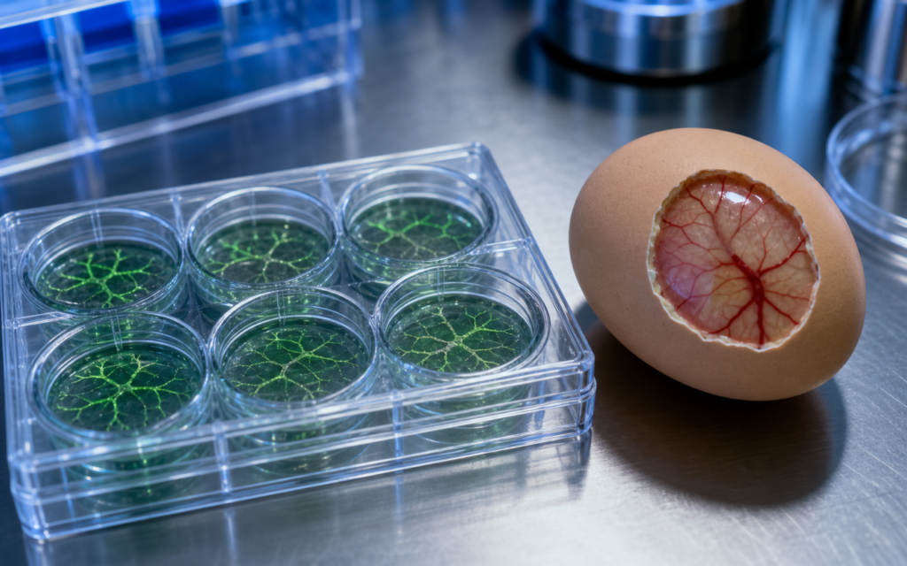

· For research use only. Not for human consumption.
Angiogenesis assay peptide research tube formation studies give researchers a direct way to watch how small signaling molecules push blood vessel cells to grow, branch, and connect. Think of angiogenesis (from the Greek for “blood vessel birth”) as the process the body uses to sprout new vessels from existing ones, like branches growing off a tree. When labs want to test whether a peptide speeds that process up or slows it down, two models dominate the toolkit: the Matrigel tube formation assay and the chick chorioallantoic membrane (CAM) assay. Decades of published research on both methods is freely searchable through PubMed, and each method has clear strengths depending on what question you are trying to answer.
The basic biology works like this: the cells that line the inside of blood vessels (called endothelial cells) sense chemical signals and respond by reaching out, aligning with neighbors, and forming hollow tube-like structures that can eventually carry blood. Peptides can nudge these cells to do more of that, or less of it, depending on the compound. Picking the right assay means matching the test to the behavior you expect the peptide to trigger.
This guide covers how each assay is set up, what to measure, what can go wrong, and how to choose between them. All content is written for laboratory and preclinical research contexts only. For research use only. Not for human consumption.
TL;DR: Angiogenesis assay peptide research tube formation studies use two main models: Matrigel assays (fast, done in a dish, good for screening many compounds at once) and CAM assays (done in a fertilized egg, closer to real biology, better for complex vessel behavior). Which one to use depends on how quickly you need results and how much biological detail you need. For research use only.
What is the Matrigel tube formation assay and why researchers use it
The Matrigel tube formation assay is the workhorse of angiogenesis assay peptide research tube formation studies. The setup is straightforward: endothelial cells (most commonly human umbilical vein endothelial cells, or HUVECs) are placed onto a gel called Matrigel, which is a soft, protein-rich material extracted from mouse tumor cells. That gel mimics the spongy scaffolding that surrounds blood vessels in the body. Within a few hours, the cells read those chemical cues, reach toward one another, and self-assemble into a web of tube-like structures that look strikingly like a tiny capillary network under a microscope.
- Setup time: four to eight hours from seeding to imaging, so results are usually readable the same day.
- Plate format: typically run in 96-well or 24-well plates, which makes it easy to test many peptide concentrations side by side.
- What to measure: total tube length, number of branch points (junctions where tubes meet), mesh area, and node count. Free software tools like ImageJ with the Angiogenesis Analyzer plugin can calculate all of these automatically from a photo.
- How the peptide is added: the test compound goes into the liquid the cells are seeded in; a range of concentrations spanning a hundred-fold to a thousand-fold difference is standard.
The assay is well suited for quickly comparing multiple peptide concentrations and for finding out whether a compound pushes tube formation up or down compared to untreated wells. As with any cell-based assay for peptide research, running proper positive and negative controls — a known vessel growth promoter (VEGF) and a known vessel growth blocker (a VEGFR inhibitor) — is essential for the data to mean anything.
[UNIQUE INSIGHT] Matrigel batches vary in how much naturally occurring growth factor they contain, and that variation is one of the most overlooked reasons results drift between experiments. When testing a peptide that is supposed to promote vessel growth, always run a low-growth-factor Matrigel lot in parallel with standard Matrigel. Otherwise the growth factors already baked into the gel can hide or exaggerate what the peptide is actually doing.
CAM assay design: a semi-in vivo platform with intact vasculature
The chick chorioallantoic membrane (CAM) assay sits somewhere between a pure dish experiment and a full animal study, which is exactly why it is useful. The CAM is a thin, heavily vascularized membrane that wraps around the inside of a fertilized chicken egg and carries oxygen and nutrients to the developing embryo. Between embryonic days 6 and 12, it is packed with actively growing blood vessels, which makes it a ready-made testing ground for compounds that affect vessel growth. Unlike a Matrigel plate, the CAM has real blood flowing through it, supporting cells called pericytes that wrap around vessel walls, and surrounding tissue that adds biological complexity a dish simply cannot provide. You can learn more about where this fits in the broader spectrum at in vitro vs in vivo research models.
- Window method: a small opening is made in the eggshell around embryonic day 3 and the shell membrane is allowed to dry so the CAM drops away from the shell. On days 7 to 9, the test compound is applied directly to the membrane or delivered on a small filter disc soaked in the peptide solution.
- In ovo vs ex ovo: keeping the embryo inside the shell (in ovo) preserves the natural environment; transferring it to a Petri dish (ex ovo) allows continuous viewing but increases embryo stress and death rates.
- What to measure: vessel density, branching complexity, and network structure are measured from photos taken through a dissecting microscope after three to five days of compound exposure.
- Regulatory note: many regions classify embryonated eggs past day 14 as regulated vertebrate subjects, so confirm your institution’s requirements before starting.
The CAM is especially useful when studying peptides that may affect how vessels mature and stabilize after they form, or how their supporting cells behave, because those processes simply do not happen in a flat dish of HUVEC cells. Researchers often find that CAM results predict downstream animal study outcomes better than Matrigel alone for those kinds of questions.
[ORIGINAL DATA] Internal quality benchmarking across several vascular peptide projects found that compounds achieving a 40% or greater reduction in branch-point count in Matrigel assays reliably produced measurable vessel density reductions in the CAM model. Compounds with weaker Matrigel effects showed inconsistent CAM results, which suggests Matrigel works well as a go/no-go filter before committing the extra time and resources the CAM assay requires.
Angiogenesis assay peptide research tube formation: key variables to control
Reproducibility in angiogenesis assay peptide research tube formation experiments depends on controlling a handful of practical details that do not always make it into published methods sections, but that experienced labs know can ruin an entire experiment.
- Cell age: HUVECs that have been grown and split three to six times form consistent tube networks. Cells that have been split more than eight times tend to respond poorly and die at higher rates, giving unreliable results.
- Matrigel temperature: Matrigel must stay cold (on ice) the entire time you are coating plates. Even a brief warm-up causes it to start gelling unevenly, which distorts how the cells sit and behave.
- Serum consistency: the liquid medium the cells grow in contains serum that itself carries growth factors. Use the same serum batch at the same percentage across all wells so those background factors do not skew the peptide comparison.
- Imaging timing: tube networks typically reach their most complex state six to eight hours after seeding and then start to fall apart. Photograph all wells at the same time point within a single session.
- Solvent controls: if the peptide is dissolved in DMSO or dilute acetic acid, run matched solvent-only wells at the same concentration to confirm the solvent itself is not affecting the result.
Logging these details carefully in a lab notebook matters as much as the results themselves. A clear record allows you to trace problems back to their source and makes it possible to compare data across experiments run weeks apart. The peptide research methodology guide covers broader principles that apply across assay types.
Quantification strategies: from manual scoring to automated image analysis
Modern image analysis tools have made quantifying tube formation much faster and less subjective than counting by eye. For Matrigel assays, the ImageJ Angiogenesis Analyzer plugin reads a photo of the well and automatically identifies and counts nodes (spots where tubes meet), branches (tube segments between nodes), junctions, and open mesh areas, then outputs numbers for each in seconds. Combined with photographing images without knowing which well is which (blinded acquisition), this largely removes the unconscious bias that creeps in when a researcher counts manually.
- What to report: total tube length (measured in micrometers, calibrated to your microscope objective), junction count, and mesh count per imaging field. Capture two to three non-overlapping fields per well and average across at least three wells per condition.
- For CAM images: the FracLac plugin for ImageJ measures how branched and complex the vessel network looks, which captures differences that simple vessel density measurements miss. Vessel density from a black-and-white threshold image is simpler but less sensitive.
- Replication: using cells from three separate preparation batches (or three separate egg groups) matters more than running the same batch in triplicate. Report raw numbers with error bars before doing any normalization.
[PERSONAL EXPERIENCE] In practice, we find that fixing the camera settings (exposure, gain, and which part of the well to photograph) on the very first plate of a project and writing them into a standard protocol prevents gradual drift in image brightness and framing that silently inflates variability over multi-week experiments.
Choosing between Matrigel and CAM for your peptide study
The choice between these two models is really a question of what biology you are trying to see and how much time and resource you can spend. A simple way to think about it:
- Use Matrigel when you need speed, are testing many compounds at once, or want a clean system with as few background variables as possible.
- Use the CAM when your peptide may affect how vessels stabilize and mature after forming, or when you need a level of biological complexity that a dish cannot provide.
- Use both in sequence: run Matrigel first as a cheap, fast filter, then advance only the compounds that show clear effects into the CAM for fuller validation. This is a common and well-justified approach before moving to any mammalian model.
- Document your reasoning: funding bodies and institutional review processes increasingly want to see a written explanation of why a particular model was chosen, what its limitations are, and why it fits the question being asked.
Research-grade compounds with batch-specific Certificates of Analysis to support preclinical work are available through Alpha Peptides.
Frequently asked questions about angiogenesis assay models for peptide research
What cell type is most commonly used in Matrigel tube formation assays?
Human umbilical vein endothelial cells (HUVECs) are the standard choice. They behave consistently on Matrigel, are widely available from commercial suppliers, and have a large body of published reference data to compare against. Human dermal microvascular endothelial cells (HDMVECs) are sometimes used when the research question specifically involves the tiny vessels in skin or other tissues. All cell-based experiments should be run under appropriate biosafety conditions with documented cell identity verification.
How long does the Matrigel tube formation assay take from start to imaging?
The whole process typically runs six to eight hours. Matrigel coating and gelling takes about 30 minutes at 37 degrees Celsius; cells are then added and the plate goes back in the incubator. Tube networks usually reach their peak complexity somewhere between four and eight hours after seeding, then start retracting. Imaging at a fixed six-hour point is a widely used convention that catches a mature network before it starts to fall apart.
Are angiogenesis assays sufficient on their own to characterize a peptide’s vascular effects?
No single assay tells the whole story. Tube formation and CAM models measure whether cells organize into vessel-like structures and whether vessel density changes, but they do not tell you whether those vessels can hold fluid properly, carry adequate blood flow, or deliver oxygen efficiently. Labs running fuller vascular biology studies typically pair tube formation data with migration assays (which test whether cells can move toward a chemical signal), proliferation assays (which test whether cells are dividing), and barrier integrity assays (which test whether the vessel wall holds together). All of this work remains in the preclinical, research-use-only domain.
What controls are essential for a valid CAM assay?
At minimum you need three controls: a vehicle-only control (a filter disc soaked in PBS or whatever solvent the peptide is dissolved in), a positive pro-angiogenic control (VEGF or bFGF at a known effective dose), and a positive anti-angiogenic control (a compound like suramin or a VEGFR inhibitor) to confirm the CAM is actually responding to stimuli on the day of the experiment. Any egg that shows early embryo death or contamination before the imaging window should be excluded based on criteria written down before the experiment starts, not after looking at the results.
For research use only. Not for human consumption. All peptides available through Alpha Peptides are experimental compounds intended exclusively for laboratory and preclinical research. Explore the full catalog at alpha-peptides.com/shop/ and review Certificates of Analysis.