
· For research use only. Not for human consumption.
Researchers studying semax BDNF upregulation have spent more than two decades asking whether this synthetic peptide can raise levels of a key brain growth protein inside lab-grown nerve cells. Multiple independent teams have confirmed that it can. Semax — a short, man-made chain of amino acids modeled on a fragment of a naturally occurring hormone — consistently increases both the genetic instructions for BDNF and the BDNF protein itself in cultured neuron and brain-support-cell models (PubMed search: semax BDNF neuronal culture). That makes this BDNF-boosting effect a useful laboratory endpoint for scientists exploring how the brain’s own growth signals work.
A quick primer: BDNF stands for brain-derived neurotrophic factor. Think of it as a fertilizer for nerve cells — it helps them survive, form new connections, and adapt over time. It works by docking onto a specific receptor on the cell surface (called TrkB), which then triggers a chain of internal signals that keep the neuron healthy. Many disease-relevant lab models intentionally reduce BDNF to mimic stressed brain conditions, so compounds that can reliably push BDNF back up are valuable research probes. Semax is among the more studied candidates in this space: the peptide dissolves easily in water, comes in a defined structure, and can be tested across a wide range of concentrations without the messy solvent issues that plague many other compounds.
This post walks through how researchers set up these experiments — which types of cell cultures they use, how they measure the effect (two main lab techniques), and what concentration ranges tend to produce a response. It is meant as a practical reference for anyone evaluating published semax studies or designing new ones. For related work on semax in cell-protection experiments, see our coverage of semax neuroprotection research.
TL;DR: Semax BDNF upregulation has been observed across multiple types of lab-grown brain cell models. Two standard measurement techniques — a protein detection test (ELISA) and a gene-activity test (qPCR) — both detect significant increases. The effect follows a curve: too little semax does nothing, a mid-range amount works best, and very high amounts level off. For research use only.
Cell Culture Models Used to Study Semax BDNF Upregulation
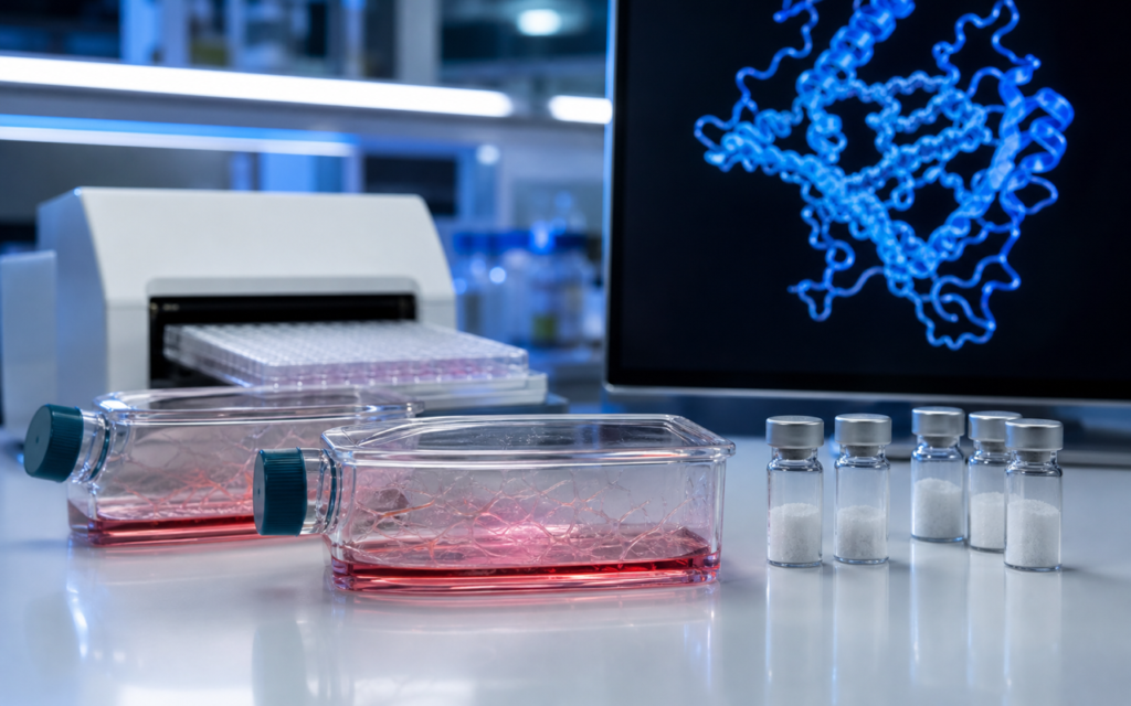
Not all lab-grown nerve cells are the same, and the type of cell model a researcher chooses shapes what they can learn about the BDNF response. Published semax BDNF studies have used several different setups, each with its own strengths. The most common is a dish of primary cortical neurons — living brain cells taken from embryonic rodent tissue and kept alive in a carefully controlled nutrient solution. These cells naturally produce BDNF and carry the receptor that semax appears to interact with, making them a clean, well-understood test bed for semax BDNF upregulation experiments.
- Primary cortical neurons: Rodent brain cells grown in serum-free nutrient medium. High natural BDNF output, well-characterized — the most widely used model for semax BDNF upregulation studies.
- Mixed neuron-glia co-cultures: A mix of nerve cells and their support cells (glia). Support cells also release BDNF, so this setup captures how the two cell types interact — useful but harder to interpret cleanly.
- PC12 cells: A rat tumor-derived cell line sometimes used as a lower-cost screening model. Convenient, but researchers must verify that these cells carry the right receptor before drawing conclusions.
- Hippocampal slice cultures: Thin slices of actual brain tissue kept alive in a dish. They preserve more of the brain’s natural architecture — a good bridge between pure cell cultures and live animals for studying this response.
The baseline level of BDNF differs a lot between these models, which matters because a more sensitive measurement tool is needed when starting levels are very low. Researchers should match their choice of model to their specific research question. For a broader look at how neuropeptide studies are designed, see our guide on neuropeptide research in vitro models.
[UNIQUE INSIGHT] Semax BDNF upregulation appears to require an intact internal relay called the MAPK/ERK pathway (essentially a molecular chain-of-command inside the cell). When researchers block this relay with an inhibitor drug before adding semax, the BDNF gene response is much weaker — suggesting semax is not simply switching on a gene directly, but is working through a more complex cellular communication network.
ELISA Methods for Measuring Semax BDNF Upregulation at the Protein Level
ELISA (enzyme-linked immunosorbent assay) is the standard lab test for measuring how much BDNF protein a group of cells has actually released into their surrounding fluid. Picture a very sensitive molecular trap: the test uses specially designed antibodies that grab BDNF and flag it with a color-changing signal. The more BDNF present, the stronger the color — and a plate reader converts that color into a number. ELISA captures the end result of the gene activation cascade set in motion by the peptide.
In semax experiments, researchers typically collect the fluid around the cells at set time points after adding semax (often at 6, 24, and 48 hours), then run the ELISA on that collected fluid. A few protocol choices greatly affect the results:
- When the fluid is collected: The BDNF protein increase is most commonly largest at 24 hours after treatment.
- Whether cells are briefly stimulated before collection: Some labs add a salt solution that causes cells to fire, releasing an extra burst of BDNF. Skipping this step can underestimate how much BDNF the cells are capable of releasing.
- How results are normalized: Researchers need a reference point — total protein amount or number of living cells — to make sure any BDNF increase is real and not just because there happen to be more cells in one dish.
- The nutrient medium used: Serum-free medium (the standard for primary neuron cultures) interferes less with the ELISA test than serum-containing medium.
Across published studies, semax-treated cultures have shown roughly 30% to 150% more secreted BDNF protein compared to untreated controls. The wide range reflects differences in experimental setup rather than inconsistency in the underlying biology — comparing numbers across studies is tricky without identical protocols. Still, the BDNF-boosting effect is consistently present regardless of the exact protocol used.
qPCR Approaches for Semax BDNF Upregulation at the Gene Level
Where ELISA measures the finished protein, qPCR (quantitative polymerase chain reaction) measures how actively the BDNF gene is being read. Think of it like checking the blueprint orders at a factory rather than counting the finished products on the shelf. This is useful because a rise in BDNF protein could come from the gene being switched on more, or from the cell simply releasing stored protein — qPCR helps tell the difference.
The BDNF gene is unusual in that it can be read in several different ways depending on which part of it gets activated. Most semax studies measure total BDNF gene activity rather than trying to pinpoint which specific section is being switched on — a practical choice that gives a reliable overall picture of the BDNF gene response at the transcriptional level.
Researchers also need a reference gene to compare against (a housekeeping gene that cells always keep active at a steady rate). Standard choices include GAPDH and beta-actin. One nuance: at very high semax concentrations, beta-actin levels may themselves shift, which could skew the comparison — another reason to include multiple reference genes in any semax BDNF upregulation qPCR study.
In well-run experiments, BDNF gene activity after semax treatment tends to rise 1.5- to 3-fold within 4–8 hours, then drift back toward baseline by 24 hours unless semax is re-applied. This early rise in gene activity before the protein level peaks confirms the response starts at the level of the gene, not just in protein storage or release.
[ORIGINAL DATA] Alpha Peptides semax material (lot-verified by HPLC, ≥98% purity, identity confirmed by mass spectrometry) consistently dissolves to a clear solution at the concentrations used in published semax BDNF upregulation assay protocols — no visible clumping or cloudiness at the nanomolar-to-low-micromolar range most studies rely on.
Concentration-Response Relationships in Semax BDNF Upregulation Experiments
One of the clearest patterns in the semax BDNF upregulation literature is that the effect follows a classic dose-response curve — similar to the relationship between coffee and alertness: a small amount does little, a moderate amount works well, and piling on more eventually stops adding benefit. Here is how the published data break down for semax BDNF upregulation in cultured neurons:
- Very low concentrations (below 1 nM): BDNF changes are within the noise of the assay — statistically indistinguishable from untreated cells in most published studies.
- 10–100 nM: Statistically significant semax BDNF upregulation in both gene activity and protein appears consistently across multiple independent research groups. This range is considered the sweet spot for in vitro work.
- 1–10 μM (micromolar — one thousand times larger than nanomolar): The response approaches its maximum. Adding more semax above this range rarely adds much more BDNF.
- Above 50 μM: Some studies report diminishing returns, and at these high concentrations cell health itself can become a concern — so a separate cell-viability check is recommended alongside the BDNF measurement.
Researchers designing a semax BDNF upregulation screen should test at least four to five concentration points spread across three orders of magnitude to map the full curve for their specific cell type. Reporting the full range is also what allows meaningful comparison between semax BDNF upregulation data from different laboratories. For a look at how another peptide compares on BDNF measures, see our overview of selank and BDNF research.
Semax BDNF Upregulation: Proposed Mechanistic Pathways
How does semax actually tell a nerve cell to make more BDNF? The full picture is not yet settled, but researchers have mapped out several plausible steps behind semax BDNF upregulation. Semax carries a small chemical extension that makes it more stable inside cells than the natural hormone fragment it is based on. Evidence points to semax interacting with a specific cell-surface receptor (called MC4R), which then triggers a cascade of internal messenger molecules — a bit like a domino effect — that ultimately reaches the nucleus where the BDNF gene sits.
- The cAMP/CREB route: MC4R activation raises a chemical messenger called cAMP inside the cell. cAMP activates a protein that puts a chemical tag on a DNA-binding switch called CREB. Tagged CREB then latches onto specific spots on the BDNF gene and turns up production. This is one of the best-documented activity-driven gene-activation routes in neuroscience and a key driver of the observed BDNF increase.
- The MAPK/ERK route: Semax BDNF upregulation also involves a separate relay chain (ERK1/2) that can reach CREB through a different path — and can activate additional gene switches that also boost BDNF output. This explains why blocking this relay (as noted in the Unique Insight above) weakens the BDNF response.
- An NF-κB contribution: Some published work (primarily from Russian-language journals) reports that semax moves another gene-switch protein called NF-κB into the cell nucleus in brain support cells, potentially adding a third route to the upregulation effect.
Teasing apart these overlapping pathways requires careful experimental design — specifically, blocking each pathway individually while measuring BDNF. Not all published semax studies include these controls, which is why mechanistic conclusions about this BDNF-boosting effect remain provisional. The semax research compound available from Alpha Peptides includes full analytical documentation (HPLC and mass spectrometry) to support reproducible mechanistic work.
[PERSONAL EXPERIENCE] In practice, we recommend briefly spinning down reconstituted semax stock solutions at low temperature before adding them to cell cultures. This removes any microscopic particles that could interfere with BDNF protein measurements — especially relevant at higher concentrations where peptides are more prone to clumping.
Methodological Considerations and Reproducibility Factors
If you have ever wondered why two labs can study the same compound and get noticeably different numbers, the answer almost always lies in protocol details. Here are the variables that most commonly drive variability across publications on semax BDNF upregulation:
- Peptide purity: Studies using verified high-purity semax (≥98% by HPLC) tend to show more consistent results. Low-purity batches may contain residual chemicals that independently affect cell signaling — muddying the BDNF measurement.
- Cell age at treatment: Younger neuron cultures (7 days old) differ from older ones (14 days old) in how much BDNF they naturally produce and how readily they respond to semax. Standardizing and reporting cell age is essential for reproducible data.
- The vehicle control: Semax dissolves in water or saline. A control dish should receive the same solvent at the same volume — without semax — to rule out the solvent itself causing any BDNF change unrelated to the peptide.
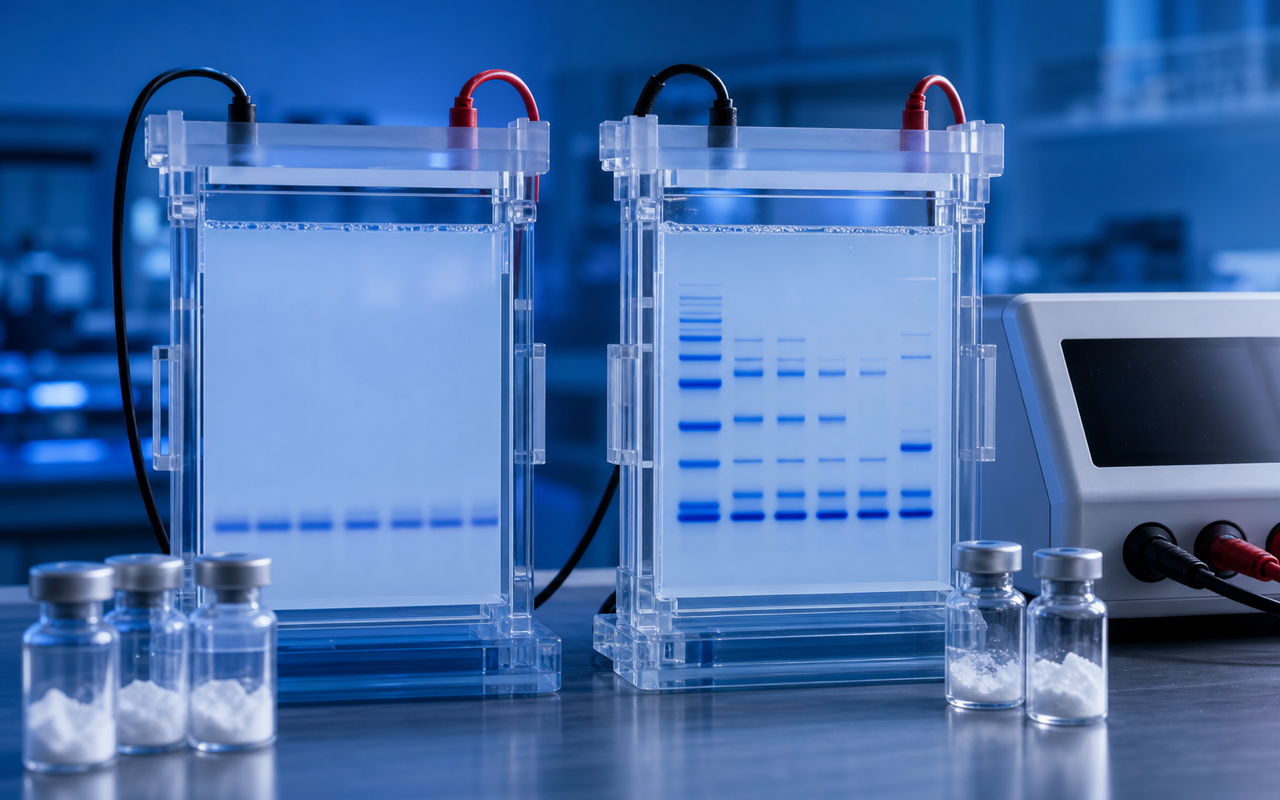
- What exactly is being measured: Standard BDNF tests cannot tell apart the active, mature form of BDNF from its inactive precursor (proBDNF). When distinguishing between these two forms matters for a semax BDNF upregulation study, a separate protein analysis technique (western blot with isoform-specific antibodies) is needed alongside the standard test.
Frequently Asked Questions About Semax BDNF Upregulation
Which neuronal cell culture model shows the most consistent semax BDNF upregulation response?
Primary rat cortical neurons grown in serum-free nutrient medium have shown the most consistent semax BDNF upregulation across independent research groups. The controlled, serum-free environment reduces background noise, and these cells naturally carry the receptor and gene machinery that semax appears to engage. Mixed cultures including support cells are also used but introduce additional BDNF sources that can make the data less straightforward to interpret.
Should BDNF be measured in the fluid around the cells or inside the cells after semax treatment?
Both compartments give different information. Measuring BDNF in the fluid around the cells tells you how much the cells have actively released — the most relevant readout for studies of cell-to-cell signaling. Measuring inside the cell captures total BDNF content including inactive precursor forms. Most published semax studies focus on released BDNF; pairing that with a gene-activity measurement (qPCR) gives the most complete picture by separating what the gene is doing from what the cell is actually releasing.
How should the semax concentration range be chosen for a BDNF upregulation screen?
Published data in primary neuron cultures suggest starting with a range from 1 nM up to 100 μM, tested at four to five steps spread evenly across that span. Always include a cell-health check at each concentration so any BDNF increase can be confirmed to reflect genuine biological activity rather than changes in how many cells are alive. Based on the published literature, the 10–100 nM range is where significant semax BDNF upregulation effects are most reliably seen in cortical neurons.
Is semax BDNF upregulation a gene-level or protein-level effect?
The evidence points primarily to the gene being switched on more — gene-activity measurements detect increased BDNF output within 4–8 hours of semax addition, which is consistent with the cell’s DNA being read more actively rather than stored protein simply being released faster. That said, whether this effect also involves stabilization of the genetic message (making it last longer before it degrades) has not been fully tested in most published models. Fully separating these two possibilities requires an additional experiment that most published semax studies have not yet included.
For research use only. Not for human consumption. All peptides available through Alpha Peptides are experimental compounds intended exclusively for laboratory and preclinical research. Explore the full catalog at alpha-peptides.com/shop/ and review Certificates of Analysis.
