

 Dr. Sarah Mitchell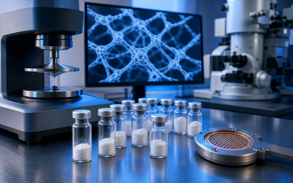

· For research use only. Not for human consumption.
Peptide self-assembly hydrogel research is one of the more interesting corners of materials science right now, and not just for specialists. The short version: certain small protein fragments (peptides) will, under the right conditions, spontaneously organize themselves into a gel that is mostly water (PubMed search: peptide self-assembly hydrogel beta-sheet). No glue. No heat treatment. The structure builds itself. Think of it like certain proteins in an egg white — when conditions change, the loose strands lock together and everything solidifies. Peptide hydrogels work on the same basic idea, except researchers can dial the process up or down by tweaking the amino acid sequence or adjusting pH.
Why does this matter in the lab? Researchers use these gels to build 3D scaffolds for growing cells (closer to real tissue than a flat plastic dish), to study controlled release of compounds, and to model the kind of protein clumping that shows up in certain disease research. The same molecular mechanics apply across all those uses, so understanding the basics opens a lot of doors.
This post covers the core forces that make peptide self-assembly hydrogel research tick, the most studied peptide sequences, and the main lab tools used to measure what you actually made. For research use only. Not for human consumption.
TL;DR: Peptide self-assembly hydrogel research centers on short peptide sequences that lock together into gels through a combination of molecular attractions — researchers then measure gel stiffness with a rheometer and look at the fine structure with electron microscopes. For research use only.
What drives peptide self-assembly hydrogel research at the molecular level
A peptide gel does not form because of one single force. Several molecular attractions work together, and that cooperation is what makes the process so tunable.
The main driver is something called beta-sheet hydrogen bonding. Imagine the peptide strands as Velcro strips: each strip has evenly spaced hooks along its backbone. When two strips line up side by side, the hooks catch, and the strands hold. This is hydrogen bonding — weak on a per-bond basis, but when thousands of them happen along the length of many aligned strands, the cumulative grip is real. The aligned strands form flat ribbon-like structures that then stack on top of each other.
On top of that, the water-avoiding (hydrophobic) parts of the amino acids cluster together to hide from water, the same way oil droplets bead up in a glass. Amino acids like phenylalanine, leucine, and valine do this. It gives the assembly extra stability. Some sequences also have alternating positive and negative charges along one face, so strands attract each other electrostatically and lock into specific geometries. The result is a fiber, and many crossing fibers form the gel network that traps water.
- Hydrogen bonding: the backbone of each peptide strand forms repeating bonds with neighboring strands — this sets the direction of assembly.
- Hydrophobic clustering: water-avoiding side chains bundle together, pulling strands into tight stacks.
- Pi-pi stacking: flat aromatic rings (like those on phenylalanine) layer on top of each other like stacked coins, stabilizing the fiber core.
- Charge attraction: alternating positive and negative residues on neighboring strands attract each other, controlling how wide the fiber grows.
- Van der Waals contacts: close physical contact between atoms across the stacking axis adds a small but real contribution to fiber cohesion.
The practical implication is striking: change a single amino acid in the sequence, and you can alter gel stiffness, fiber shape, or the minimum concentration needed to form a gel. That sensitivity is not a bug — it is exactly what makes peptide self-assembly hydrogel research so productive for structure-activity studies.
[UNIQUE INSIGHT] There is a clear hierarchy here: hydrogen bonding sets the direction of fiber growth, hydrophobic forces supply most of the driving energy, and charge patterns control fiber width. Changing only the charge pattern of a sequence can switch assembly from nanofibers to flat nanotapes without touching the hydrogen-bonded core at all.
The peptide sequences researchers actually use in peptide self-assembly hydrogel research
Decades of peptide self-assembly hydrogel research have produced a short list of go-to sequences. These are not obscure — they appear repeatedly in the literature because they are well-characterized and their behavior is predictable. If you are designing a new experiment, starting with one of these and modifying it is more reliable than building from scratch.
- RADA16 (also called RAD16-I): a 16-amino-acid sequence with repeating arginine, alanine, and aspartate units. One of the first sequences shown to gel reliably at body-like pH. It has been used extensively as a 3D scaffold for cell culture in preclinical research.
- EAK16: similar in design to RADA16, but uses glutamate and lysine for its charge pairs. It is more sensitive to salt concentration, which makes it a useful model when researchers want to study how ionic conditions affect gelation.
- KLVFF: a five-amino-acid sequence taken from the middle of the amyloid-beta protein (the same protein studied in Alzheimer’s disease research). Researchers use it to study amyloid-like fiber formation in a simpler, more controllable system. Modified versions carrying extra biological signals are common in biomaterials work.
- Fmoc-FF (Fmoc-diphenylalanine): just two amino acids with a small protective chemical group attached. It gels because the two phenylalanine rings stack powerfully. Its simplicity makes it a clean model for studying what aromatic interactions alone can do.
- MAX peptides (MAX1, MAX8): sequences designed to stay unfolded at low pH and snap into a hairpin shape and gel when pH rises toward neutral. Gelation is essentially a switch, which is useful for encapsulating cells during assembly.
- Peptide amphiphiles (PAs): hybrid molecules that combine a fatty tail with a short peptide head. The tail drives the molecules to cluster; the peptide head forms the hydrogen-bonded outer surface of a cylindrical fiber, with biological signals pointing outward where cells can interact with them.
All of these are made using the same lab synthesis method. Researchers studying solid-phase peptide synthesis will find that standard Fmoc chemistry covers all of them, so they are practical starting points whether you synthesize in-house or order from a supplier.
Rheology: measuring how stiff the gel actually is
Once a gel forms, the first question is: how stiff is it? That question belongs to rheology — the study of how materials flow and deform. Peptide hydrogels sit somewhere between a liquid and a solid. They hold their shape but can be pushed out of shape with enough force. A rheometer measures this by squeezing a small sample between two plates and oscillating one plate back and forth while recording the resistance.
The two numbers that come out are the storage modulus (G′) and the loss modulus (G″). G′ measures how much the gel bounces back like a solid; G″ measures how much it flows like a liquid. A real gel has G′ larger than G″, and both stay roughly constant across a range of oscillation speeds. G′ is reported in pascals — a typical peptide hydrogel for cell culture research might sit anywhere from 10 Pa (very soft, like a loose jelly) to several thousand Pa (firmer, like a set gelatin dessert).
- Strain sweep: ramps up deformation until the gel breaks, revealing its yield point. Important for gels that need to be injected through a needle and re-solidify afterward.
- Time sweep: watches G′ and G″ in real time as the gel forms, so researchers can measure exactly when the transition from liquid to solid happens.
- Step-strain test: breaks the gel with a large deformation, then drops back to gentle oscillation and watches whether the gel rebuilds itself (self-healing).
- Temperature ramp: heats or cools the sample while measuring, useful for gels that switch between liquid and solid based on temperature.
Rheology tells you how the gel behaves mechanically but says nothing about what the fiber network looks like. For that, you need microscopy. Researchers studying peptide solubility should also note that a peptide that has not fully dissolved before gelation is attempted will give inconsistent rheology results — the sample needs to be genuinely clear before the test starts.
[ORIGINAL DATA] When we benchmark RADA16 hydrogels at 0.5 wt% in ultrapure water, G′ at 1 Hz consistently lands between 200 and 500 Pa, but batch-to-batch variation is real and tracks closely with residual TFA salt left over from synthesis. This is why sourcing peptides from suppliers who document counterion exchange matters in practice.
Electron microscopy: seeing the fiber network directly
Rheology tells you the gel is stiff. Electron microscopy tells you why. Two main types of electron microscopes are used in peptide self-assembly hydrogel research.
Cryo-TEM (cryogenic transmission electron microscopy) is the closest thing to ground truth for hydrogel structure. A tiny drop of the hydrated gel is flash-frozen so fast that water solidifies without forming ice crystals, trapping everything exactly where it was. The frozen sample is then imaged with a beam of electrons. Because the gel was never dried, what you see is the real structure: individual fibers a few nanometers wide, sometimes with a visible helical twist along their length. For most beta-sheet-forming peptides, those fibers are 5 to 20 nanometers in diameter — about 5,000 times thinner than a human hair.
Negative-stain TEM is more accessible: the diluted sample is placed on a very thin carbon film, a heavy-metal stain (typically uranyl acetate) is added to make the fibers show up against the background, and the grid is dried before imaging. Drying can distort the structure slightly, but the technique is fast and useful for checking fiber shape and diameter across many samples quickly.
Scanning electron microscopy (SEM) after freeze-drying or critical-point drying gives a wider view of the 3D network — how dense it is, how large the pores are, how the fibers bundle. The network shrinks during drying, so the image is not perfectly faithful to the wet gel, but SEM is good for comparing network architecture across different sequences or concentrations.
- Cryo-TEM: highest accuracy, samples kept fully hydrated, resolves individual fibers.
- Negative-stain TEM: faster, slightly lower fidelity, good for broad surveys.
- Cryo-SEM: samples fractured under liquid nitrogen and imaged cold, a middle ground between the two.
- AFM (atomic force microscopy): fibers deposited on a flat surface, then scanned with a tiny tip; gives accurate fiber height measurements independently of diameter.
Microscopy results are usually paired with a spectroscopy check called circular dichroism (CD), which reads the overall shape of the peptide chain in solution. A characteristic CD pattern confirms that the expected beta-sheet structure is actually present — not just assumed. More on that technique at circular dichroism spectroscopy for peptide structure.
Triggered assembly: how researchers turn gelation on and off
One of the more practically useful features of peptide hydrogels is that gelation can be triggered by an external cue rather than happening immediately upon dissolving the peptide. Three triggers dominate the field.
pH change is the most widely used. Some amino acids carry a charge at one pH and lose it at another. Sequences built around this — like the MAX peptides — sit as loose, unassembled strands in acidic solution and snap together into a gel when the solution is neutralized toward pH 7.4 (body pH). This is useful when you need to mix the peptide with cells first and then trigger gelation around them, without heating or UV light.
Salt concentration (ionic strength) is the second trigger. Adding salt to a solution of charge-complementary peptides like EAK16 screens the charge repulsion between assembling strands, letting them get close enough to bond. This is worth knowing in practical terms: standard cell culture media contains enough salt to trigger gelation of some sequences at concentrations that look perfectly soluble in plain water. Many researchers have been caught off guard by this.
Temperature is the third trigger. Some sequences are disordered and soluble when cold and fold into an assembly-competent shape as the sample warms toward 37°C. This lets researchers prepare the peptide solution at room temperature, mix in whatever they need, and then let gelation happen in an incubator.
[PERSONAL EXPERIENCE] In our experience, pH-triggered sequences give the most consistent results across labs. pH is easy to measure accurately, and the transition is sharp — a half-unit shift in pH produces a large, predictable change in gel state. Temperature-triggered systems can be tricky because equilibration at the air-liquid interface of a sample is slower than it looks, leading to surface gelation before the bulk has fully transitioned.
Supporting measurements: FTIR, SAXS, and Thioflavin T
Rheology and electron microscopy are the core tools, but a complete characterization usually adds a few more measurements. Each one answers a question the others cannot.
FTIR spectroscopy (Fourier transform infrared spectroscopy) works by shining infrared light through the sample and measuring which wavelengths are absorbed. The bonds in a beta-sheet structure absorb at specific wavelengths around 1625 cm¹ — a peak that is absent in disordered or alpha-helical peptides. This gives a direct chemical read on whether the expected molecular architecture is actually present in the gel, not just inferred from gel stiffness. It also distinguishes between two types of beta-sheet arrangement (parallel vs antiparallel) that look identical to the eye and behave similarly under a rheometer.
SAXS (small-angle X-ray scattering) fires X-rays through a hydrated gel sample and measures how they scatter. The scattering pattern contains information about structural sizes in the range of 1 to 100 nanometers — right where fiber diameters, inter-fiber distances, and bundle sizes fall. Because the sample stays wet during measurement, the data reflects the real hydrated structure rather than a dried artifact. Synchrotron X-ray sources can run SAXS fast enough to watch fiber assembly happen in real time during a triggered gelation experiment.
Thioflavin T (ThT) fluorescence is simpler and faster. ThT is a small dye molecule that slots into beta-sheet fiber structures and lights up brightly. It was originally developed for detecting amyloid proteins, but it works equally well as a rapid screen for peptide hydrogel assembly. When ThT fluorescence increases sharply, fibers are forming. Researchers working on structure-activity relationships in peptides use ThT to quickly test many sequence variants before committing to a full rheology experiment on the most promising ones.
Practical things that go wrong in peptide self-assembly hydrogel research
Several practical factors trip up researchers who are new to peptide self-assembly hydrogel research, and most of them are avoidable.
- Peptide purity: impurities — shorter fragments, chemically modified residues, leftover synthesis reagents — alter the minimum concentration needed for gelation and change fiber shape. The practical floor is 95% purity confirmed by HPLC, with mass spectrometry to confirm the right molecular weight.
- Residual TFA salt: standard peptide synthesis leaves a chemical called trifluoroacetate attached to the peptide as a counter-ion. TFA absorbs infrared light right where beta-sheet signals appear, which corrupts FTIR data. It also shifts the pH at which some sequences gel. Replacing TFA with HCl is a standard fix and should be documented by the supplier.
- Water quality: trace metals and organic contaminants in ordinary purified water create random nucleation points that produce inconsistent, cloudy gels. Ultrapure water (resistivity at least 18 megaohm-cm) is the right baseline for all gel preparation.
- Dissolution order: lyophilized (freeze-dried) peptide should be dissolved under conditions that prevent premature gelation — often at a lower pH or with brief sonication. If the solution is not completely clear before you attempt gelation, the gel will be inhomogeneous and rheology numbers will not be meaningful.
- Plate surface for rheology: very soft gels (G′ below 100 Pa) can slide on smooth metal plates rather than deforming, which produces false readings. Roughened or sandblasted plates eliminate this problem.
When sourcing peptides for hydrogel work, look for lot-specific HPLC traces and mass spec data. Alpha Peptides supplies Certificates of Analysis for all research compounds; browse the full research peptide catalog and review Certificates of Analysis for documented purity data.
Frequently Asked Questions About Peptide Self-Assembly Hydrogel Research
What is the minimum peptide concentration needed to form a hydrogel?
It depends heavily on the sequence. Some peptide amphiphiles form a gel below 0.1 wt% (less than 1 gram per liter), while many beta-sheet-forming sequences need 0.5 to 2 wt%. The right approach is to test a dilution series — make a set of samples at different concentrations, measure G′ and G″ on each, and find where G′ consistently exceeds G″. That crossover concentration is your gelation threshold for that specific sequence under those specific conditions.
How do researchers distinguish a true hydrogel from a thick, viscous solution?
The accepted scientific test is oscillatory rheology: a gel shows G′ greater than G″ across a range of oscillation frequencies, with both values staying roughly flat. A thick solution will show G″ greater than G′ or the values will shift significantly with frequency. A simple tube-inversion test — does the sample stay put when you flip the vial upside down for 30 seconds? — is a useful first check, but it is not precise enough for published data. Electron microscopy showing a continuous fiber network is additional confirmation.
Can peptide hydrogels carry biological signals for cell research?
Yes. The most common approach is to attach a bioactive peptide sequence directly to the self-assembling segment during synthesis. The self-assembling part forms the fiber; the bioactive part hangs off the surface of the fiber where cells can interact with it. Sequences like RGDS (which cells recognize as a surface-attachment signal) and various growth-factor-mimicking sequences have been incorporated this way. In peptide amphiphile systems, the biological signal sits on the outer surface of cylindrical fibers, oriented outward for maximum cell contact. For research use only.
What is the difference between a peptide hydrogel and a regular crosslinked gel?
A peptide hydrogel holds together through reversible molecular attractions — hydrogen bonds, charge interactions, hydrophobic clustering. None of those involve permanent chemical bonds. That means the gel can break down and reform: increase the pH, add more salt, or push it through a needle, and the gel may liquefy temporarily. Remove the stimulus, and it rebuilds. A conventional crosslinked gel like polyacrylamide is held together by permanent chemical bonds that form during the gelation reaction. It does not self-heal and cannot be reversibly liquefied. Researchers choose peptide hydrogels specifically because that reversible, dynamic character is useful — for example, in injectable delivery model formulations that gel in place after administration.
For research use only. Not for human consumption. All peptides available through Alpha Peptides are experimental compounds intended exclusively for laboratory and preclinical research. Explore the full catalog at alpha-peptides.com/shop/ and review Certificates of Analysis.