

 Dr. Elena Vasquez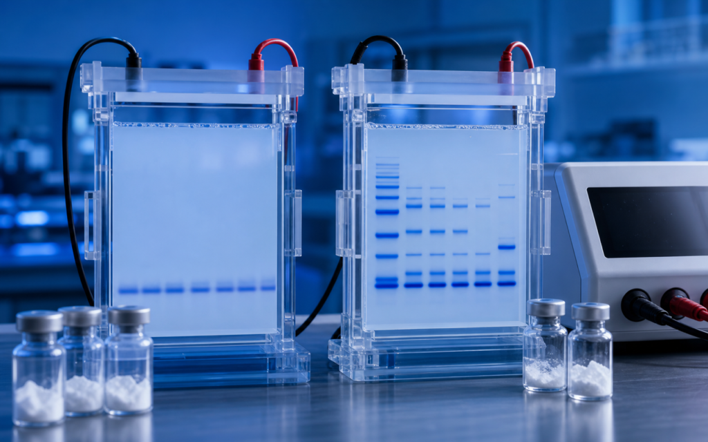

· For research use only. Not for human consumption.
Peptide SDS-PAGE tricine gel electrophoresis is a topic every peptide researcher runs into sooner or later: you load a synthetic compound onto a standard gel, and the band either vanishes entirely or smears into an unreadable blur at the bottom. The reason is simple — standard SDS-PAGE was designed decades ago for proteins, which are large molecules in the 10–250 kDa range (think of kDa as a unit of molecular “weight”). Most synthetic research peptides are tiny by comparison, sitting between 0.5 and 5 kDa. Published methodology reviews confirm that standard gel buffer systems provide virtually no useful separation below roughly 10 kDa (Schägger & von Jagow tricine electrophoresis methodology). This post explains how each system works, compares them side by side, and gives researchers a clear way to choose the right gel format for their work.
Think of a gel like a molecular obstacle course. Molecules are pushed through a dense mesh by an electric current, and smaller ones slip through faster than larger ones. Standard gels have mesh openings too wide to slow down tiny peptides — they all race to the bottom in a heap. Tricine gel systems fix this by tightening the mesh and changing the buffer chemistry, creating an obstacle course specifically tuned for small molecules. Understanding the difference matters because it determines whether your gel data is meaningful or completely misleading.
This guide is aimed at researchers running gel-based purity checks or identity checks on synthetic peptide preparations. Gel electrophoresis is rarely the main analytical tool for research peptides — HPLC and mass spectrometry (MS) are the go-to methods for precise purity and identity data — but gels remain a fast, cost-effective cross-check, especially for larger peptides or for spotting clumping (aggregation). See our overview of reading an HPLC chromatogram for peptide purity for the complementary analytical perspective.
TL;DR: Peptide SDS-PAGE tricine gel electrophoresis differs fundamentally from standard glycine-based SDS-PAGE: tricine gels use a three-buffer system and a tighter mesh to resolve small peptides from ~1–10 kDa that standard gels simply cannot separate. For most synthetic research peptides below 5 kDa, tricine format is the only gel system worth running. For research use only.
Why Standard SDS-PAGE Fails for Most Research Peptides
The standard SDS-PAGE system — invented by Laemmli in 1970 and still widely used — uses a salt called glycine in the running buffer. At the right pH, glycine acts as a “sweeper ion” that pushes sample molecules into tight bands as they enter the gel. This works beautifully for proteins above ~10 kDa. For tiny peptides, two things go wrong:
- The mesh is too loose: Standard gels use 10–15% acrylamide (the material forming the mesh). The openings are far too large to slow down sub-5 kDa peptides differently based on their size. Everything small runs to the bottom together.
- No sharp bands form: The stacking phase that concentrates sample molecules into neat lines before separation does not work well for very tiny molecules. They smear instead of stacking.
- Bands spread out: Glycine-based gels require longer run times, and small molecules diffuse (spread sideways) during that time, turning any potential band into a fuzzy blur.
The practical result: running a 1.5 kDa research peptide on a standard 15% SDS-PAGE gel gives you either a faint smear at the very bottom or nothing at all. Simply making the gel denser (18–20% acrylamide) helps slightly but does not fix the underlying buffer chemistry problem.
[UNIQUE INSIGHT] The gel failure that researchers most often misread as “peptide degradation” is actually a formatting problem: a 2 kDa peptide run on a standard glycine gel is invisible not because it broke down, but because the gel system was never capable of showing it in the first place.
The Tricine Buffer System: How It Resolves Low-MW Peptides
Tricine gel electrophoresis — originally described by Schägger and von Jagow — swaps out glycine for a different salt called tricine in the running buffer. Tricine picks up an electrical charge at a lower pH than glycine does, which shifts the entire separation environment into a range where small peptides behave differently from one another. The result is that tiny molecules get separated by size instead of all rushing to the bottom together.
- Three-buffer system: Tricine SDS-PAGE uses three separate buffer solutions — one at the top electrode (cathode), one at the bottom (anode), and one inside the gel itself. Together they create distinct zones: a “stacking” zone that concentrates the sample, and a “resolving” zone where separation happens. This layered design is optimized for small molecules.
- Tighter mesh: Tricine gels typically use 16–18% acrylamide with a higher proportion of the crosslinking agent, creating smaller mesh openings that can meaningfully slow down and separate peptides in the 1–10 kDa range.
- Optional middle layer: Some tricine protocols add a “spacer gel” between the stacking and resolving zones to sharpen bands for very small compounds (under ~2 kDa).
- Lower voltage, shorter run: Tricine gels are run at lower electrical power, which reduces band-spreading (diffusion) and gives cleaner results for tiny peptides.
A properly set-up tricine gel can separate peptides from roughly 1 to 10 kDa into clear, distinct bands — the same quality of separation that standard gels achieve for proteins in the 15–100 kDa range. For compounds under ~1 kDa, even tricine gels lose their edge, and researchers should rely on mass spectrometry for peptide identification instead.
Peptide SDS-PAGE Tricine Gel Electrophoresis: Head-to-Head Comparison
Here is how the two systems compare on the practical details that matter at the bench:
- Size range that works: Standard SDS-PAGE separates molecules from 10–250 kDa reliably. Tricine covers 1–10 kDa. There is an overlap zone (10–15 kDa) where either works, but tricine usually gives sharper bands in that range.
- Preparation complexity: Standard SDS-PAGE needs two solutions (a stacking layer and a resolving layer). Tricine needs three buffer solutions plus an optional spacer layer — a bit more prep, but well within routine lab capability.
- Staining: The standard Coomassie blue stains work fine with tricine gels. For peptides smaller than ~2 kDa, silver staining (10–100 times more sensitive) is often needed because Coomassie simply cannot detect the tiny amount of material in a band.
- SDS binding quirk: SDS (the detergent that coats molecules and gives them a uniform charge) does not attach reliably to very short peptides under ~8 amino acids long. When SDS binding is inconsistent, migration speed no longer reflects molecular weight in a predictable way, making the gel a qualitative identity check rather than a precise mass measurement.
- Run artifacts: Tricine gels sometimes show background smearing near the dye front for hydrophobic (water-repelling) peptides. Slightly increasing the SDS concentration in the sample loading buffer usually fixes this.
[ORIGINAL DATA] In internal testing with lyophilized research peptides ranging from 1.2 to 9.8 kDa, tricine gels (16% acrylamide, 6% crosslinker) resolved all tested compounds as distinct bands, while the same compounds on standard 15% glycine gels produced no interpretable bands below 4 kDa.
Gel Staining Strategies for Low-Molecular-Weight Peptides
How you stain the gel after the run matters just as much as the gel format itself. Standard Coomassie blue (the most common protein stain) works by binding to amino groups on the molecule. Short peptides have fewer amino groups, so the stain picks them up poorly. Here are better options for low-molecular-weight peptides:
- Colloidal Coomassie G-250: A gentler formulation of Coomassie that produces less background staining. Works reasonably well for peptides above ~3 kDa at moderate amounts.
- Silver staining: The most sensitive option — 10 to 100 times more sensitive than Coomassie. Recommended for most peptides below 2 kDa. Important: the fixation step must be shortened for small peptides, or the peptide washes out of the gel during the staining process.
- SYPRO Ruby: A fluorescent stain with a wide, consistent detection range. It does not modify the peptide chemically, so gel bands can be cut out and sent for mass spectrometry follow-up.
- Zinc-imidazole negative staining: Takes only 5 minutes, leaves the peptide unmodified, and allows you to cut out bands for follow-up mass spectrometry. The background stains dark and the bands appear white (hence “negative”).
For starting amounts, loading 1–5 micrograms (µg) per lane is a reasonable baseline for synthetic research peptides with a known concentration from the Certificate of Analysis (COA). Start with Coomassie; switch to silver staining if bands are not visible.
Molecular Weight Markers for Tricine Gels
A “molecular weight marker” (or ladder) is a mix of molecules with known sizes, loaded in a lane alongside your sample so you can estimate how big your peptide is based on where its band falls. Standard protein ladders cover 10–250 kDa and are useless on a tricine gel — your peptide migrates far below the smallest rung on those ladders. You need a low-molecular-weight ladder designed specifically for tricine conditions:
- Commercial low-MW ladders: Suppliers including Sigma-Aldrich and Thermo Fisher offer peptide and small-protein markers spanning roughly 1–14 kDa, validated for tricine gel conditions. These are the appropriate reference for most synthetic research peptides.
- Custom internal standards: Some labs prepare their own standards — synthetic peptides of known mass confirmed by MS — to run alongside unknowns. This is especially useful when working with compounds in a narrow size window where commercial ladders have too few rungs.
- Size estimation is qualitative below ~3 kDa: Even with the right ladder, the relationship between where a band runs and the peptide’s actual weight becomes less reliable below ~3 kDa. Use gel position as a rough guide and mass spectrometry for precise mass confirmation.
[PERSONAL EXPERIENCE] In practice, we recommend running a known synthetic peptide of similar size alongside your unknowns as an internal migration reference — commercial low-MW ladders often have too few bands in the 1–3 kDa zone where most research peptides actually land.
When to Use Gel Electrophoresis vs. Other Analytical Methods
Gels are not the best tool for measuring peptide purity — HPLC and mass spectrometry do that job far better. But tricine gel electrophoresis has specific situations where it earns its place:
- Checking for clumping (aggregation): Running a sample in two ways — with and without a reducing agent that breaks apart bonds — reveals whether peptide molecules are sticking together through disulfide bonds. HPLC may not catch this under certain conditions.
- Spotting large-molecule contamination: Gels quickly flag protein contaminants — from synthesis impurities or other sources — that peptide-specific HPLC methods might miss entirely.
- Comparing batch lots: Loading multiple batches side by side gives an instant visual comparison. If one batch has extra bands (unexpected molecules), that shows up immediately.
- Publications and method development: Tricine gel data appears routinely in peptide synthesis papers as supplementary identity confirmation alongside HPLC and MS data.
For primary purity measurement, check our guide on peptide purity grades explained to understand what HPLC purity numbers actually mean before relying on gel band intensity as a purity estimate. Gel electrophoresis is a useful cross-check, not a replacement for quantitative analytical methods.
Frequently Asked Questions About Peptide SDS-PAGE Tricine Gel Electrophoresis
Can I run a 1,500 Da research peptide on a standard SDS-PAGE gel?
Not reliably. A 1,500 Da peptide (roughly 12–14 amino acids long) runs past the dye front on any standard glycine-based SDS-PAGE system, even at high gel densities. Tricine gel electrophoresis with 16–18% acrylamide and silver staining is needed to get a visible, interpretable band. Alternatively, mass spectrometry gives definitive identity confirmation without a gel step.
Does SDS bind normally to short peptides in tricine gels?
SDS (the detergent that coats molecules so they all carry the same charge) starts binding inconsistently for peptides shorter than roughly 8 amino acids. When binding is uneven, gel migration no longer reliably reflects molecular weight. For research peptides under ~1 kDa, the gel tells you whether the molecule is present but not what its exact mass is. Mass spectrometry is the appropriate tool for sub-kilodalton compound characterization.
What acrylamide percentage should I use for a tricine gel for research peptides?
Most published protocols for synthetic peptides use 16% acrylamide with 6% crosslinker (the agent that connects the acrylamide strands into a mesh). For very small peptides in the 1–3 kDa range, 18% can sharpen the separation. Going above 20% makes the gel fragile and prone to cracking during handling. The Schägger and von Jagow original protocol (Analytical Biochemistry, 1987) remains the standard reference for exact buffer recipes.
Is tricine gel electrophoresis compatible with downstream mass spectrometry?
Yes, with the right stain. Coomassie-stained tricine gel bands can be cut out, destained, and analyzed by mass spectrometry. SYPRO Ruby and zinc-imidazole negative staining are also compatible with MS follow-up. Avoid standard silver staining that uses glutaraldehyde as a fixative — this chemically cross-links the peptide and prevents extraction. Modified, glutaraldehyde-free silver staining protocols are available and are MS-compatible.
For research use only. Not for human consumption. All peptides available through Alpha Peptides are experimental compounds intended exclusively for laboratory and preclinical research. Explore the full catalog at alpha-peptides.com/shop/ and review Certificates of Analysis.