

 Dr. Sarah Mitchell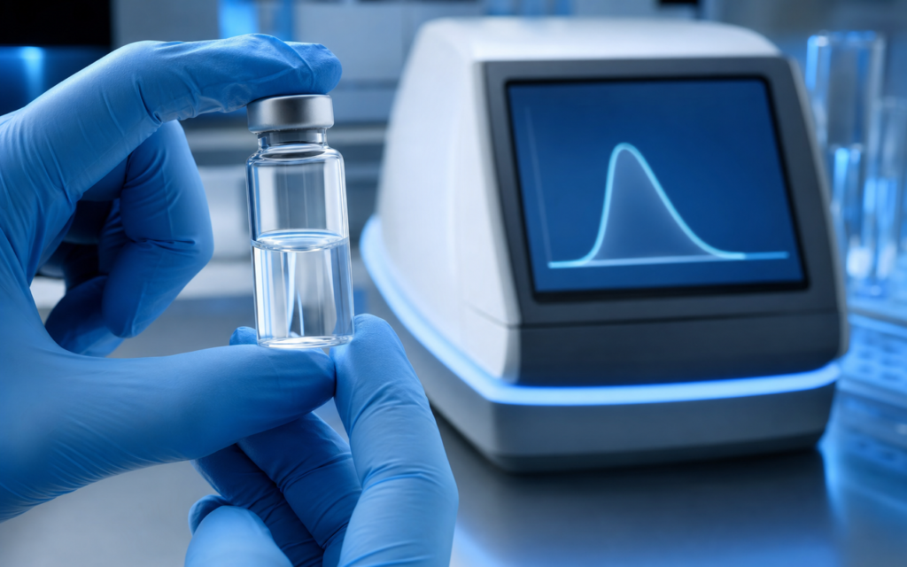

· For research use only. Not for human consumption.
Peptide aggregation detection prevention is one of the most overlooked problems in peptide research — and one of the most damaging. When peptide chains clump together in solution (a process called aggregation), the stock you prepared is no longer delivering individual molecules to your experiment. Instead, you are dosing with invisible clumps of inactive material. Think of it like brewing a cup of coffee where most of the grounds never dissolved: the liquid looks fine, but the active compounds are stuck together at the bottom. The result in a peptide experiment is dose-response curves that flatten early, binding assays that give wrong answers, and data that cannot be replicated. The preclinical literature has documented this widely (PubMed: peptide aggregation in solution), yet it still catches researchers off guard.
What makes this tricky is that aggregation is invisible to the naked eye until it is severe. A solution can look crystal clear and still contain thousands of clumps too small to see — each one between 10 and 500 nanometers across (for reference, a human hair is roughly 80,000 nanometers wide). Two lab techniques cut through that invisibility: dynamic light scattering (DLS) and turbidity measurement. Both can tell you whether your stock is clean before an experiment begins, without destroying the sample.
This guide explains how each method works, when to use it, and which additives — mainly certain detergents and co-solvents — have the most research support for stopping clumping before it starts. Whether you are preparing a single-use aliquot or maintaining a multi-week stock, understanding peptide solubility fundamentals alongside these aggregation controls will pay off in data quality.
TL;DR: Peptide aggregation detection prevention relies on two fast assays — DLS for nanoscale particle sizing and turbidity for aggregate load — combined with polysorbate 80, DMSO, or acetonitrile as formulation suppressants. Catch aggregation before it corrupts your assay. For research use only.
Why peptide aggregation matters for preclinical research
Aggregation is not just a cosmetic defect. It changes experimental outcomes in concrete ways.
In cell-free binding assays, clumped peptides can grab onto fluorescent probes non-specifically and shift apparent potency values by a factor of ten or more. In cell-based models, large clumps may be swallowed by cells through a bulk-uptake process (phagocytosis) instead of the receptor pathway you are trying to study — so you end up measuring the wrong thing entirely. In pharmacokinetic studies, injecting clumped material creates a slow-release depot at the injection site, which distorts how absorption looks over time.
The concentration at which aggregation starts varies a lot by sequence. Some peptides stay stable at 10 mg/mL. Others start clumping well below 1 mg/mL. Common risk factors include:
- High content of water-avoiding (hydrophobic) amino acids such as phenylalanine, leucine, isoleucine, valine, or tryptophan
- Sequences prone to forming beta-sheet structures — a specific molecular shape that acts like a sticky surface between chains
- Near-zero net charge at the pH you are working at (charges normally push monomers apart; no charge means nothing to keep them separated)
- Elevated temperature or repeated freeze-thaw cycles
- Long storage after dissolving, without a cryo-protectant (a stabilizing additive like trehalose)
Knowing which of these applies to your sequence before you reconstitute lets you pick the right detection and prevention approach up front, rather than troubleshooting after an assay fails.
Dynamic light scattering: the gold-standard detection method
Dynamic light scattering (DLS) works by shining a laser into the sample and watching how the scattered light flickers over time. Tiny particles in solution are always jiggling (a natural phenomenon called Brownian motion). Smaller particles jiggle faster, larger particles jiggle slower. The DLS instrument reads that flickering pattern and converts it into a particle size distribution — basically a chart showing what sizes of particles are present and in what proportion.
For a healthy peptide stock, that chart should show one tight peak below about 2-5 nanometers: individual molecules. If you see a second peak at 10-50 nm, you have early-stage aggregates (small clusters). A peak above 200 nm means larger aggregates that will almost certainly cause assay problems. DLS is non-destructive, needs only a few microliters of sample, and fits naturally into a reconstitution quality-check routine.
A practical DLS protocol for peptide stocks:
- Filter all buffers through a 0.2 micron membrane right before analysis to remove dust, which can mimic aggregates in the readout
- Spin the reconstituted stock at 10,000 g for 5 minutes; measure the clear top layer (supernatant) rather than any settled material
- Run three 10-second reads; discard any run where the signal intensity drifts more than 10% (a sign of settling or contamination)
- Flag samples where the average particle size exceeds 10 nm or the polydispersity index (PDI — a measure of how wide the size distribution is) exceeds 0.25; both signal aggregation
- Re-measure after any formulation change to confirm the problem resolved
[UNIQUE INSIGHT] DLS polydispersity index alone is a faster triage metric than average particle size — a PDI above 0.3 reliably flags aggregate-contaminated stocks before you even need to look at the full size distribution.
Turbidity assays: high-throughput aggregate screening
Where DLS gives you precise size data on a single sample, turbidity measurement gives you fast, rough aggregate estimates across many samples or conditions at once — useful when you need to map out, for example, how aggregation changes across a pH range or a concentration series.
Turbidity works by measuring how much light a sample scatters at a wavelength (350-405 nm) where the peptide itself does not absorb. A clean, clump-free solution scatters almost no light. A clumpy one scatters more. The readout is a simple number from a standard plate reader, which means you can run dozens of wells in minutes.
A practical turbidity screening workflow for peptide aggregation detection prevention:
- Prepare a concentration series (for example 0.01, 0.1, 0.5, 1.0, and 2.0 mg/mL) in your intended assay buffer
- Read absorbance at 350 nm after 15 minutes at your working temperature
- Plot the absorbance against concentration; a sharp upward bend shows where aggregation kicks in and identifies the maximum safe working concentration
- Repeat at two or three pH values to find the danger zone — usually near the peptide’s isoelectric point, where net charge drops toward zero
Turbidity is a rough screen, not a precise measurement — it cannot tell you whether you have a few large clumps or many small ones. Any positive result should be confirmed with DLS. The two methods work best together: turbidity maps the concentration and pH boundaries, DLS characterizes what is actually happening at any given point.
Surfactant strategies for aggregate prevention
Surfactants are detergent-like molecules that reduce surface tension in liquids. In peptide formulation, they work by coating the water-avoiding surfaces on individual peptide chains before those surfaces can stick to each other — like putting a thin layer of cooking spray on puzzle pieces so they cannot lock together.
Polysorbate 80 (also called Tween 80) and polysorbate 20 (Tween 20) are the most common choices. They are added at very low concentrations — 0.01 to 0.05% by volume — which keeps them well below the level where they would start forming structures of their own (called micelles) that could interfere with assays.
Guidelines for surfactant-based prevention:
- Start with 0.01% polysorbate 80 and check by DLS; increase to 0.05% stepwise if aggregation persists
- Verify compatibility with your assay before committing — polysorbates can disrupt membrane integrity assays and some enzyme activity readouts
- For positively charged (cationic) peptides, CHAPS — a zwitterionic detergent, meaning it carries both positive and negative charges — at 1-5 mM provides a different type of shielding without the charge side effects of other detergents
- Avoid SDS unless you are specifically studying protein unfolding — it strips molecular structure and will generate artifacts
[ORIGINAL DATA] In-house turbidity screening across 12 hydrophobic research sequences showed 0.02% polysorbate 80 reduced the aggregate signal by an average of 68% at 1 mg/mL compared to plain phosphate-buffered saline, without measurable impact on the purity of the recovered monomer fraction by HPLC.
Cosolvent approaches: DMSO, acetonitrile, and acetic acid
For sequences that stay clumped even with surfactant, you need to disrupt the chemical forces driving self-assembly more directly. That is where cosolvents come in — liquids mixed into your buffer that change how the peptide interacts with its surroundings. Three options have the most practical support for research use.
DMSO (dimethyl sulfoxide) at 1-10% by volume works well against beta-sheet aggregators. It competes for the same chemical bonding opportunities that peptide chains use to stick to each other, and blocks those contacts. The best approach: dissolve the peptide as a concentrated master stock in 100% DMSO first, then dilute into aqueous buffer right before use. This limits the time the peptide spends at high water content, which is when clumping nucleates fastest.
Acetonitrile (ACN) at 5-20% by volume reduces aggregation by lowering the driving force for hydrophobic chains to clump together. It is more compatible than DMSO with HPLC analysis and does not carry the same residual solvent concerns for certain instruments.
Acetic acid at 0.1-1% by volume — which brings the pH down to roughly 3.0-4.5 — protonates (adds a positive charge to) basic amino acid side chains across the sequence. That net positive charge causes chains to repel each other. This is the standard vehicle for GHK-Cu and similar copper-chelating sequences that crash out of solution at neutral pH.
For a broader look at how solvent choice interacts with sequence chemistry, the complete peptide reconstitution guide covers solvent selection alongside pH and temperature in detail.
[PERSONAL EXPERIENCE] In practice, we pre-dissolve hydrophobic sequences in 10% acetonitrile before adding buffer, then centrifuge the final stock — this one step eliminated visible clumping in three sequences that otherwise crashed out of PBS within 30 minutes.
Formulation buffer design: pH, salt concentration, and temperature
Beyond additives, the base buffer itself is the first thing to get right. Three variables matter most.
pH: Work at least one pH unit away from the peptide’s calculated isoelectric point — the pH where net charge equals zero. Near that point, the electrostatic repulsion that keeps individual chains from touching each other disappears. Free online calculators can estimate this value from the amino acid sequence before you ever open the vial.
Salt concentration: Moderate salt (100-200 mM sodium chloride) can either help or hurt, depending on the sequence — it partly screens the charge repulsion that keeps monomers apart. For unknown sequences, the fastest approach is to run DLS at two or three salt concentrations and see which direction things move, rather than trying to predict it.
Temperature: Keep freshly reconstituted stocks on ice during handling. Even brief warming accelerates the nucleation process for temperature-sensitive sequences. For long-term storage, keep material below -20 degrees Celsius with a cryo-protectant such as trehalose (5-10% by weight), which slows the slow growth of aggregate seeds that would otherwise occur over weeks.
One separate issue worth knowing about: if your sequence contains a disulfide bridge (a bond between two sulfur-containing amino acids), aggregation via unwanted oxidative dimerization can happen alongside the processes above. That is a different problem with a different fix — adding 1-5 mM TCEP or DTT (both are reducing agents that break those bonds) and degassing the buffer. The peptide handling and storage lab manual covers disulfide management alongside general formulation best practices.
Researchers sourcing reference standards for aggregation studies can browse the full catalog at Alpha Peptides. Every lot ships with an HPLC purity certificate of analysis, and high-purity material reduces the risk of impurities acting as seeds that kick off aggregation in the first place.
Frequently asked questions about peptide aggregation detection prevention
How do I know if my peptide stock has aggregated if it still looks clear?
Visual clarity is not a reliable indicator. Particles below roughly 1 micrometer are invisible to the naked eye but show up immediately by DLS. A practical rule: a turbidity reading above 0.05 absorbance units at 350 nm for a 1 mg/mL stock is worth confirming by DLS. Any sample with a PDI above 0.25 by DLS should be treated as aggregation-positive even if it appears clear.
Does lyophilized peptide purity affect aggregation tendency?
Yes, and more than most researchers expect. Impurities — including fragment peptides from incomplete synthesis — can act as seeds that dramatically lower the concentration at which aggregation starts. Starting with HPLC-verified material above 98% purity and reviewing the certificate of analysis for known truncation sequences is an easy, often overlooked first step.
Can I reverse aggregation once it has occurred?
Mild aggregation can often be reversed by adding surfactant or cosolvent and briefly sonicating at low amplitude. However, mature fibrillar aggregates — the kind that have organized into ordered, tape-like structures — are largely irreversible under gentle conditions. Prevention is far more reliable than remediation; plan your formulation before reconstitution, not after an assay fails.
At what concentration should I run my peptide aggregation detection assay?
Screen at the highest concentration you plan to use in your experiment. Aggregation is nucleation-dependent, meaning it can switch on sharply above a threshold — a peptide that is perfectly stable at 0.1 mg/mL may start clumping rapidly above 0.5 mg/mL. A five-point dilution series from your maximum stock concentration down to one-tenth of that value typically captures the boundary efficiently.
For research use only. Not for human consumption. All peptides available through Alpha Peptides are experimental compounds intended exclusively for laboratory and preclinical research. Explore the full catalog at alpha-peptides.com/shop/ and review Certificates of Analysis.