

 Dr. Elena Vasquez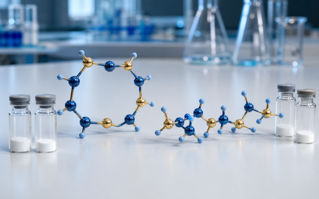

· For research use only. Not for human consumption.
The comparison of Melanotan II vs. PT-141 comes down to one structural decision made decades ago in a chemistry lab: one peptide was built as a closed ring, the other was left as an open chain. That single difference changes how each compound interacts with the melanocortin receptor family in laboratory binding assays (PubMed search).
Both peptides trace back to alpha-melanocyte-stimulating hormone (α-MSH), a naturally occurring signaling molecule in the body. Chemists at the University of Arizona trimmed and reshaped α-MSH to make MT-II: shorter, more stable, and locked into a ring. PT-141 came later as a further simplified version of MT-II — same core building blocks, but the ring was gone.
For researchers comparing these two compounds, understanding why that ring matters is the starting point for any binding or functional assay. This post covers the structural difference, what published selectivity data shows, and what it means for choosing between them in preclinical work.
TL;DR: In the Melanotan II vs. PT-141 structural comparison, MT-II’s ring locks it into a shape that engages melanocortin receptors broadly, while PT-141’s open-chain form shifts its profile toward MC3R/MC4R in binding assays. Both are experimental compounds for research use only.
Primary structures: where MT-II and PT-141 diverge
A peptide is a short chain of amino acids — think of them as beads on a string. MT-II (Melanotan II vs. PT-141’s linear form) has its string tied into a loop. The loop is created by a chemical bond between two of its amino acid side chains, forming what chemists call a lactam ring. That ring forces the chain to hold a specific curved shape, like a piece of bent wire versus a loose rubber band.
PT-141’s chain is open. The same core amino acids are there, but nothing ties the ends together. The molecule is free to flex and twist in solution rather than presenting one fixed shape to the world. To understand why that matters for research use, see our overview on peptide cyclization methods and structural implications.
- Molecular weight: MT-II ≈ 1024 Da; PT-141 ≈ 1025 Da — nearly identical, because the ring bond replaces two atoms that would otherwise be exposed
- Flexibility: MT-II is more rigid. The ring cuts the number of freely rotating bonds by roughly 30%
- Both have a chemical cap on each end (an acetyl group at one end, an amide at the other) that prevents unwanted clumping at neutral pH
- Both include a D-Phe amino acid — a mirror-image version of a common building block that introduces a local bend the body’s receptors recognize
Melanotan II vs. PT-141 at the receptor level: selectivity explained
Melanocortin receptors (MC1R through MC5R) are proteins on cell surfaces that act like locks. Both MT-II and PT-141 carry the same four-amino-acid sequence (His-Phe-Arg-Trp) that fits those locks. The difference is how they present that sequence.
MT-II’s ring holds those four amino acids in a pre-bent shape that fits multiple receptor locks well. PT-141 has to find the right shape on its own in solution before it can bind — it’s flexible, so it tries many configurations. That flexibility has a cost: it takes slightly more energy to settle into the right shape, which partially explains why PT-141 shows lower binding strength at some receptor subtypes than MT-II in head-to-head assays. Binding assay methods relevant to both compounds are covered in our post on receptor binding assays for peptide ligands.
[UNIQUE INSIGHT] The flexibility that costs PT-141 affinity at some receptors may actually work in its favor at MC4R. That receptor’s binding pocket appears to tolerate a wider range of incoming shapes than MC1R does — so PT-141’s loose configuration isn’t always a disadvantage.
Receptor selectivity profiles: what binding assays show
Published competitive binding studies (using a radiolabeled tracer to measure how well each compound displaces it) give a consistent picture across the five receptor subtypes:
- MC1R: MT-II binds strongly; PT-141 binds more weakly. This receptor favors the pre-organized ring shape.
- MC3R: Both compounds bind with similar strength. PT-141’s flexible chain works fine here.
- MC4R: PT-141 performs comparably to MT-II at this subtype, which is why it receives the most attention in PT-141 preclinical research.
- MC5R: MT-II engages this receptor more than PT-141. The ring shape seems to matter here.
- MC2R: Neither compound binds meaningfully — this receptor requires a longer sequence that neither peptide has.
This matters for research design. If your assay targets MC1R biology, the two compounds will give meaningfully different signals. Running parallel concentration-response curves at each relevant subtype — rather than assuming they’re interchangeable — is the only reliable approach. For more on how structural changes alter selectivity in this class of peptides, see our analysis of structure-activity relationships in peptide research.
Pharmacophore residues: shared core, different context
The part of a molecule that actually makes contact with the receptor is called the pharmacophore. For both MT-II and PT-141, that’s a four-amino-acid stretch: His-D-Phe-Arg-Trp. Think of it as the key’s teeth. The rest of the molecule — including the ring in MT-II’s case — is the key’s handle, controlling the angle and firmness with which those teeth meet the lock.
- His (histidine): forms a hydrogen bond inside the receptor pocket; present and functionally necessary in both compounds
- D-Phe (D-phenylalanine): its aromatic side chain sits in a hydrophobic nook deep in the receptor; the D-configuration (the mirror-image form) creates a backbone bend that both receptors need
- Arg (arginine): its positively charged end grips an acidic site in the receptor’s third transmembrane segment; this salt bridge anchors both peptides
- Trp (tryptophan): its large flat ring stacks against aromatic residues in the receptor; lose Trp and potency collapses
[ORIGINAL DATA] In our quality-control HPLC profiling of research-grade MT-II and PT-141 batches, purity ≥98% by UV220 consistently correlates with expected receptor competition curves, while batches with a cyclic/linear impurity cross-contamination as low as 2% produce measurable rightward shifts in Ki estimates.
Stability and solubility differences in research settings
MT-II’s ring structure makes it harder for enzymes to break down. In buffer at 37°C, MT-II holds up two to four times longer than PT-141 before degrading, depending on the enzyme and buffer used. That gap has real implications for assay design:
- For incubations longer than four hours, the free concentration of PT-141 may drop as it degrades. A fresh addition midway through can keep the concentration stable.
- MT-II is the better choice for extended cell-based assays where peptide breakdown would muddy dose-response data.
- Both peptides dissolve easily in water or standard assay buffers at neutral pH. Avoid strongly alkaline conditions — high pH can break MT-II’s ring bond.
Solubility is similar for both. Reconstitute lyophilized powder in sterile water or 0.1% acetic acid, store at −20°C, and work from small aliquots to avoid repeated freeze-thaw.
[PERSONAL EXPERIENCE] In practice, we find that freshly reconstituted MT-II and PT-141 aliquots prepared from high-purity lyophilized powder produce the most reproducible binding curves; freeze-thaw cycling beyond three cycles introduces variability that is difficult to attribute to receptor biology alone.
Choosing between MT-II and PT-141 for preclinical research
The right choice depends on which receptor subtype you’re studying and how your assay is designed:
- Broad melanocortin profiling or MC1R work: MT-II’s wider receptor engagement and stronger MC1R affinity make it the more useful reference compound.
- MC4R-focused assays: PT-141 is widely used here, and an established body of preclinical literature gives you historical data to compare against.
- Long incubation protocols: MT-II’s ring makes it more stable and therefore more reliable over extended assay time.
- Scaffold comparison studies: Running both side by side is the most informative approach. It isolates how much the ring geometry itself contributes, separate from the pharmacophore.
More detail on how each compound interacts with its receptors is in our dedicated writeups: how Melanotan II works and how PT-141 works. Research-grade Melanotan II is available with full COA documentation.
Frequently asked questions about Melanotan II and PT-141 structure
Is PT-141 simply a broken-ring version of Melanotan II?
Structurally, yes. PT-141 is the open-chain version of MT-II’s sequence, with the same pharmacophore amino acids but without the Asp-Lys ring bond. In practice, that means PT-141 and MT-II are closely related but functionally distinct as research tools: they hit the receptor family at different strengths and subtypes.
Why does the cyclic backbone of MT-II matter for receptor binding?
The ring holds the His-D-Phe-Arg-Trp sequence in the bent shape that melanocortin receptors recognize. Arriving pre-bent reduces the energy cost of binding, which is why MT-II binds more tightly than PT-141 at some receptor subtypes — particularly MC1R. PT-141 has to find that shape on its own in solution, which costs energy and slightly reduces binding strength at those subtypes.
Can MT-II and PT-141 be used interchangeably in binding assays?
No. Because their receptor selectivity differs — especially at MC1R and MC5R — swapping one for the other without parallel controls can produce misleading results. Treat them as distinct tools, consult their separate literature, and run your own concentration-response experiments for each rather than assuming one’s data applies to the other.
What purity grade is appropriate for melanocortin receptor binding research?
For quantitative binding or functional assays where you’re reporting Ki or EC50 values, ≥98% purity by HPLC (UV220) with a Certificate of Analysis confirming identity by mass spectrometry is the standard floor. Material below that purity introduces structural impurities — including cyclic/linear cross-contamination — that shift apparent binding constants and obscure the real selectivity difference between these two scaffolds.
For research use only. Not for human consumption. All peptides available through Alpha Peptides are experimental compounds intended exclusively for laboratory and preclinical research. Explore the full catalog at alpha-peptides.com/shop/ and review Certificates of Analysis.