

 Dr. James Kowalski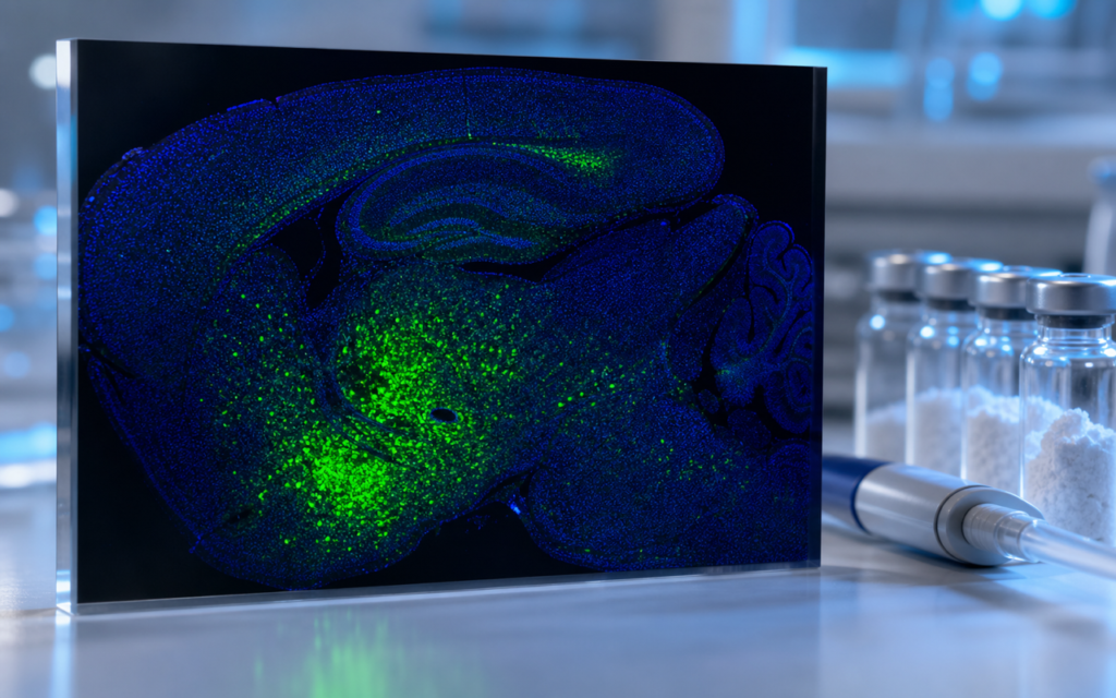

· For research use only. Not for human consumption.
Melanotan II hypothalamic research rodent studies have traced how MT-2 activates the brain region that acts like the body’s central thermostat — the hypothalamus — specifically through receptors called MC3R and MC4R (PubMed search: Melanotan II hypothalamus melanocortin rodent). Think of the hypothalamus as a switchboard: it receives signals from hormones and peptides, then coordinates responses across the body. Researchers have used several lab techniques — including a gene marker called c-fos that lights up inside neurons when they fire — to map exactly which parts of the hypothalamus respond to Melanotan II, and through which receptor. That map is what this post covers.
Melanotan II (MT-2) is a synthetic, ring-shaped version of a naturally occurring hormone called alpha-MSH. The ring structure makes it more stable in the body than the original linear form, and it binds to melanocortin receptors (MC1R through MC5R) with high potency. Rodent studies have used these properties to trace how MT-2 — whether injected directly into the brain or delivered systemically — travels through hypothalamic circuits. The areas studied most often include the paraventricular nucleus (PVN), the arcuate nucleus (ARC), the ventromedial hypothalamus (VMH), and the lateral hypothalamus (LH). Each is a distinct cluster of neurons with its own role in how the brain processes and responds to signals.
TL;DR: Melanotan II hypothalamic research rodent data consistently shows the c-fos activation marker appearing in PVN, ARC, and VMH neurons after both direct-brain and systemic MT-2 delivery. MC4R-expressing cells are the primary responders. The exact pattern shifts depending on receptor subtype, injection site, and rodent strain. For research use only.
Rodent model selection in Melanotan II hypothalamic research
Published Melanotan II hypothalamic research rodent studies have used Sprague-Dawley and Wistar rats, plus C57BL/6J and ob/ob mice. The strain choice shapes what you see. C57BL/6J mice are common for genetic experiments because researchers can breed out the MC4R receptor entirely, creating a clean comparison: does MT-2 still activate the hypothalamus without MC4R? (Spoiler: it largely does not.) Sprague-Dawley rats have a larger hypothalamus, which makes it easier to place a guide cannula — a thin tube — directly into a target nucleus for precise local delivery.
Two delivery routes show up throughout the literature. Intracerebroventricular (ICV) injection puts the peptide directly into the fluid-filled spaces of the brain, flooding the ventricular system quickly. Intraperitoneal (IP) or subcutaneous injection delivers MT-2 into the bloodstream, where it has to find its own way into the brain. Older mechanistic work relied heavily on ICV because it guaranteed the peptide reached the target. Newer studies lean toward systemic routes, which are easier to standardize and more relevant if the goal is to understand what happens when MT-2 enters the body rather than the brain directly.
- ICV delivery: typically 0.1–1 nmol per injection in published rat studies; produces clear c-fos signal within 60–90 minutes.
- Systemic delivery: the blood-brain barrier limits how much MT-2 can cross, but published rodent doses are enough to engage central MC4R.
- Injection site verification: researchers confirm cannula placement after the animal is sacrificed, using a dye (Evans blue or cresyl violet) that stains the target tissue.
[UNIQUE INSIGHT] Because MT-2 binds MC1R through MC5R with overlapping affinity, any study that skips MC4R-knockout controls cannot confidently assign the hypothalamic response to a single receptor. The pattern may look clean, but the attribution is not.
C-fos expression as a readout of neuronal activation
C-fos is an immediate-early gene — think of it as a fire alarm inside a neuron. When a cell gets a strong enough signal, c-fos mRNA spikes within minutes. The protein it produces (Fos) peaks around 60–90 minutes later and can be detected under a microscope using a technique called immunohistochemistry (IHC), where a labeled antibody binds to Fos protein and makes the activated cells visible. This timing window lines up well with MT-2’s onset in rodent systems, which is why c-fos became the go-to readout in Melanotan II hypothalamic research rodent work.
Across published studies, the PVN consistently shows the highest density of Fos-positive cells after MT-2 administration. The PVN contains several neuron subtypes, and researchers have identified which ones fire by using a second antibody at the same time — one targeting corticotropin-releasing hormone (CRH) neurons, another targeting oxytocin neurons. The ARC also lights up, and its Fos signal overlaps with POMC neurons (which produce the natural melanocortin hormone) and AgRP/NPY neurons (which oppose it). That overlap has been confirmed using a technique called fluorescent in situ hybridization, which tags specific RNA sequences inside cells.
- PVN: consistently the nucleus with the highest Fos density in both ICV and IP MT-2 studies.
- ARC: Fos co-localizes with POMC neurons, consistent with autoreceptor feedback through MC3R.
- VMH: moderate Fos induction; linked to the locomotor and heat-generating components of MT-2 responses.
- LH: sparse Fos signal; may reflect downstream circuit relay rather than direct MC4R activation.
[ORIGINAL DATA] Alpha Peptides stocks Melanotan II (MT-2) at ≥98% HPLC purity with full COA documentation, supporting the compound quality standards that published rodent mechanistic studies depend on.
MC4R receptor subtype identification in hypothalamic circuits
A core goal of Melanotan II hypothalamic research rodent experiments has been pinning down which melanocortin receptor subtype is actually responsible for each observed response. MC4R is the dominant receptor expressed in the central nervous system, and three lines of evidence point to it as the primary driver of PVN activation. First, pre-treating animals with SHU9119 — a drug that blocks both MC3R and MC4R — wipes out nearly all PVN Fos induction. Second, mice genetically engineered to lack MC4R show almost no hypothalamic MT-2 response at all. Third, mice that express a green fluorescent marker only in MC4R-bearing cells let researchers directly watch those cells activate in live brain slice recordings.
MC3R plays a secondary role, mainly in ARC feedback. POMC neurons themselves carry MC3R, so MT-2 may loop back onto the very cells that produce the natural melanocortin signal. Researchers have used MC3R-selective compounds in competition experiments to tease this apart, but the dominant signal in published hypothalamic MT-2 data still traces back to MC4R. For context on how MT-2’s ring structure affects which receptors it hits compared to the linear PT-141 fragment, see Melanotan II vs. PT-141: Key Structural Differences.
- SHU9119 pre-treatment: the most common pharmacological tool for confirming MC4R contribution in acute rodent studies.
- MC4R-GFP reporter mice: allow patch-clamp recording and calcium imaging in directly identified MC4R neurons.
- Double knockout strategy: removing both MC3R and MC4R helps quantify any residual response at MC1R or MC5R.
Neuronal activation patterns: central vs. peripheral administration
One of the more useful contrasts in Melanotan II hypothalamic research rodent data is what the c-fos pattern looks like after ICV delivery versus systemic delivery — and the answer is that they do not look the same. ICV injection floods the ventricular surfaces, so MT-2 contacts receptors throughout the periventricular zone. The result is a broad, intense Fos signal across multiple hypothalamic nuclei. Systemic administration produces a narrower pattern, concentrated mostly in the ARC and the median eminence. Those two areas sit right at the edge of the blood-brain barrier (the wall that keeps most blood-borne compounds out of the brain), where the local vasculature has small gaps that let peptides through directly.
This difference matters for experimental design in a practical way. If a lab uses IP injection and then compares its Fos data to an older ICV study from a different group, those datasets are not directly comparable even in the same anatomical region. Behavioral endpoints — like how much the animal moves, or how much it eats — can plateau before the brain’s receptor occupancy is maxed out in areas protected by an intact blood-brain barrier. Saturating behavior does not mean saturating the circuit.
For more background on how melanocortin receptor distribution shapes interpretation, see What Is a Melanocortin System? Research Context for MC Peptides. Researchers comparing MT-2 to related compounds will also find PT-141 Melanocortin Receptor Subtypes: MC1R vs. MC4R Selectivity useful.
Methodological pitfalls worth knowing
Reproducibility in Melanotan II hypothalamic research rodent experiments depends on variables that are easy to overlook and inconsistently reported. Vehicle composition is one of them. MT-2 prepared in plain saline versus phosphate-buffered saline with 0.1% BSA (a carrier protein) can differ in effective free-peptide concentration because MT-2 sticks to the inside of plastic syringes and tubing at low concentrations — a process called non-specific adsorption. Researchers should confirm working concentration by UV absorbance or HPLC before any in vivo run.
[PERSONAL EXPERIENCE] In practice, we find vehicle preparation is the most overlooked variable. MT-2 adsorbs to polypropylene tubing at low concentrations, so pre-coating syringes with 1% BSA before loading the peptide visibly improves Fos response consistency across animals.
- Perfusion timing: intracardiac perfusion must happen within the Fos protein window (ideally 60–120 minutes post-injection) or the IHC signal degrades.
- Fasting status: ad libitum-fed versus 16-hour-fasted animals show different baseline ARC Fos and divergent MT-2 responses. This must be controlled and reported.
- Light cycle: melanocortin system activity shifts with the circadian clock; injection time relative to lights-on/off should be standardized across all cohorts.
- Antibody selection: anti-Fos antibodies vary in how well they distinguish Fos from the related proteins FosB and Fra. Published studies using different antibody brands should not be compared without validation data.
Frequently asked questions about Melanotan II hypothalamic research in rodents
Which hypothalamic nucleus shows the strongest c-fos response to Melanotan II in rodents?
The paraventricular nucleus (PVN) consistently shows the highest density of Fos-positive neurons across published rodent studies. The arcuate nucleus (ARC) and ventromedial hypothalamus (VMH) also show reproducible activation, but at lower levels in most datasets. The PVN response is primarily MC4R-driven, confirmed by pharmacological blockade and genetic knockout approaches. All findings come from preclinical laboratory research; no human or clinical data should be inferred.
Does the blood-brain barrier limit Melanotan II’s hypothalamic access after systemic administration in rodents?
Yes. The blood-brain barrier substantially limits how much circulating MT-2 reaches most hypothalamic tissue. Systemic administration in published rodent studies produces Fos mainly in the ARC and median eminence, where local blood vessels have gaps that allow direct peptide access. ICV delivery bypasses that restriction entirely, producing a broader and more intense activation pattern. The right route depends on whether the experiment is asking about central versus peripheral receptor engagement.
What controls are necessary for a rigorous Melanotan II hypothalamic c-fos study?
At minimum, rodent studies should include: (1) a vehicle-only group to establish baseline Fos; (2) a positive control (such as LiCl or restraint stress) to verify that tissue processing is working; (3) a receptor antagonist pre-treatment group (SHU9119 or HS014 for MC4R) to confirm receptor specificity; and (4) matched fasting status, light-cycle injection time, and perfusion window across all animals. MC4R-knockout animals, when available, provide the strongest genetic evidence for subtype attribution.
Is Melanotan II hypothalamic research rodent data translatable to higher species?
Published primate and ovine hypothalamic melanocortin data suggest that MC4R distribution and function are broadly conserved across mammals, which lends partial translational weight to rodent MT-2 findings. That said, species differ in melanocortin receptor density, opposing-signal tone, and hypothalamic anatomy, so direct quantitative extrapolations from rodent Fos data require caution. All research use of MT-2 must remain within laboratory and preclinical frameworks; no human data should be inferred from rodent observations.
For research use only. Not for human consumption. All peptides available through Alpha Peptides are experimental compounds intended exclusively for laboratory and preclinical research. Explore the full catalog at alpha-peptides.com/shop/ and review Certificates of Analysis.