

 Dr. James Kowalski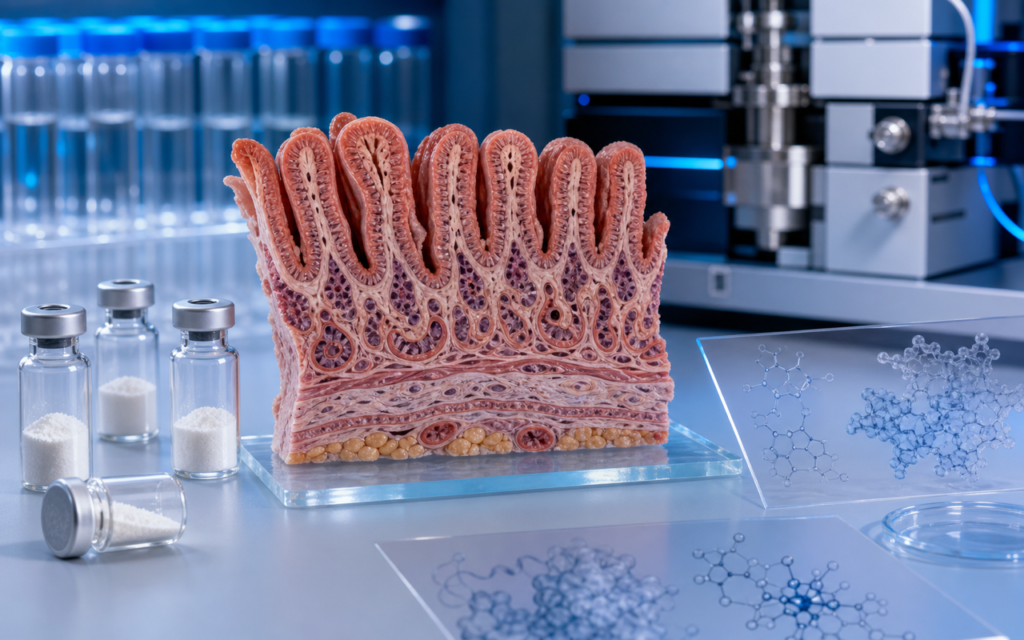

· For research use only. Not for human consumption.
GLP-2 receptor tissue expression tells researchers which body tissues actually carry the docking site for GLP-2 signals — and getting that map right is what separates a well-designed preclinical study from one that produces no useful data (PubMed search: GLP-2 receptor tissue distribution). Think of GLP2R (the GLP-2 receptor protein) as a lock, and GLP-2 as the key. If the cells in your model do not carry that lock, the key has nothing to open — and you will see no biological response no matter how well everything else is set up. Scientists first identified and cloned the gene for this receptor from rat gut tissue in the mid-1990s. At the time, the assumption was that GLP2R lived almost exclusively in the gut. More detailed mapping work done since then, using techniques that let researchers look at gene activity cell by cell rather than in whole-tissue chunks, revealed the receptor in a much wider range of locations than anyone initially expected.
This overview covers what rodent and human studies have found about where GLP2R is genuinely abundant, where it shows up at low levels, and where it is essentially absent. It is written as an orientation for researchers choosing cell or animal models, not as clinical guidance. For background on how GLP-2 compares to related peptides, see GLP-1 vs GLP-2 vs GLP-3: What’s the Difference? and What Is GLP-2?
All discussion below applies strictly to preclinical and laboratory research contexts. For research use only. Not for human consumption.
TL;DR: GLP-2 receptor tissue expression is highest in the small intestine — specifically in two cell types found there — with lower-level signals in gut neurons, certain liver cells, and a few brain regions. The most common mistake in GLP-2 research is picking a model that does not actually carry the receptor at meaningful levels. For research use only.
Why GLP-2 receptor tissue expression varies so widely
GLP2R is a receptor protein embedded in the outer membrane of certain cells. Like all proteins, it is built from instructions stored in DNA and then copied into a messenger molecule called mRNA before being assembled. The amount of receptor any given cell produces depends on how actively it reads those instructions — and that activity level varies enormously from one tissue to the next, driven by molecular switches in the DNA itself.
One practical consequence of this: broad whole-tissue measurements of GLP2R can be misleading. A tissue might test as moderately positive overall, but that signal may come entirely from one rare cell type scattered through the tissue. The surrounding cells could be producing essentially no receptor at all. A researcher who picks a cell line derived from that tissue and assumes the receptor is present may be in for a frustrating series of non-results.
There are at least three sources of variation worth knowing about:
- Gene activity differs by cell type: certain molecular switches that turn on GLP2R production are active in intestinal cells but largely inactive in liver cells, which is part of why expression is so much higher in the gut.
- The receptor gene can be read in slightly different ways, producing variant forms of the protein with different activity levels. At least two such variants have been found in rodents.
- Human and rodent GLP2R are similar — about 82% identical at the amino acid level — but not identical. Antibodies and peptides validated in one species should be re-confirmed before use in the other.
Intestinal epithelium: the primary site of GLP-2 receptor tissue expression
The small intestine carries the highest and most well-documented GLP-2 receptor tissue expression of any organ. Signal is strongest in the upper portion (duodenum and jejunum) and fades substantially by the time you reach the colon. But within the small intestine, not all cell types are equal.
Modern single-cell gene-activity studies — which measure what each individual cell is doing rather than averaging across millions of cells at once — consistently show that receptor levels are highest in two specific populations: enteroendocrine L-cells (hormone-secreting cells scattered through the gut lining) and myofibroblasts (support cells that sit just beneath the gut lining, separating it from deeper tissue layers). The absorptive cells that line most of the intestinal surface, the ones you might assume are the main players, actually carry comparatively little GLP2R.
This matters a lot for lab model design. Mini-gut organoids (tiny lab-grown replicas of intestinal tissue) built mainly from absorptive cells may show weak or no GLP-2 response, not because GLP-2 does not work in the gut, but because the model is missing the cell types that actually carry the receptor. Researchers using GLP-2 analog (glp-2-tz) in organoid assays should confirm that their preparation includes both L-cells and the myofibroblast layer before drawing conclusions from the results.
- Duodenum: high GLP2R, concentrated in myofibroblasts and scattered hormone-secreting cells.
- Jejunum and ileum: high GLP2R, consistent with the strong gut-lining growth responses seen in rodent studies.
- Colon: substantially lower GLP2R; GLP-2 responses measured here are weaker accordingly.
- Stomach lining: trace or undetectable GLP2R in most assays; not a useful primary model for GLP-2 research.
[UNIQUE INSIGHT] Single-cell gene-activity data show GLP2R is concentrated in the myofibroblast support layer beneath the gut lining, not in the absorptive surface cells. Earlier whole-tissue studies that lumped all cell types together overstated how much of the GLP-2 signal goes through the surface layer and missed how much runs through that underlying support tissue.
Enteric and central nervous system expression
The gut has its own dedicated nervous system — a dense network of neurons woven through the intestinal wall, sometimes called the “second brain” — and GLP2R turns up there too. Studies in rodents have found the receptor in gut neurons that help control local muscle movement and blood flow through the intestinal wall.
Beyond the gut, GLP2R gene activity has been detected in specific regions of the rodent brain, including parts of the hypothalamus (which regulates energy balance) and the hippocampus (involved in memory and learning). This brain-level expression is based mainly on gene-activity measurements rather than direct protein detection, so researchers should treat it as provisional and verify with additional methods in their own labs before building experiments around it. For broader background on how receptors work, see Receptor Binding: The Lock-and-Key Model Explained.
- Gut neurons: confirmed GLP2R expression, with functional evidence that GLP-2 influences gut nervous system signaling in rodents.
- Hypothalamus: GLP2R gene activity detected; possible relevance to energy-sensing circuits, studied in preclinical models only.
- Hippocampus: low-level GLP2R gene activity in rodents; what this means functionally is still under investigation.
Hepatic and vascular expression
The liver has been a source of debate. Early studies measuring GLP2R across whole liver tissue suggested a moderate signal, but more refined analyses that separate out individual cell types tell a different story. The signal appears to come mainly from stellate cells (liver cells involved in tissue repair and scarring) and cells lining the tiny blood vessels running through the liver, not from the main working liver cells (hepatocytes). Standard lab preparations of hepatocytes typically show weak GLP-2 responses, which fits with that picture.
Cells in the blood vessels supplying the intestine have also been reported to carry low but detectable GLP2R levels in rodent models. This is relevant for researchers studying how GLP-2 affects intestinal blood flow, though this area has received much less study than gut epithelial expression. Anyone designing assays around GLP-2 signaling in vascular cells should read the primary literature carefully first, because the picture is not yet fully mapped.
[ORIGINAL DATA] COA-verified GLP-2 analog purity above 98% by HPLC is important for receptor-binding studies. Small fragments of broken-down peptide can attach to GLP2R with lower binding strength and blunt the apparent potency of the intact peptide, which distorts the dose-response curve in cell-based assays.
Choosing the right in vitro model based on receptor distribution
Once you have a clear picture of GLP-2 receptor tissue expression, choosing a lab model becomes more straightforward. Here is a practical summary of common options:
- Primary small-intestinal organoids from the duodenum or jejunum: the best chance of retaining the cell types that actually carry GLP2R. Still worth confirming receptor presence by gene-expression measurement before running functional assays.
- Co-cultures that include the myofibroblast support layer: add back a GLP2R-rich population that purely epithelial preparations typically exclude.
- Caco-2 cells (a standard intestinal cell line): generally carry little or no GLP2R, because they resemble the absorptive surface cells rather than the L-cells and myofibroblasts where the receptor concentrates. Not recommended for GLP-2 receptor work without engineering them to express the receptor artificially.
- HEK293 overexpression cells: useful for studying basic receptor pharmacology, but they lack the tissue-specific signaling context that makes results from them transfer well to real intestinal biology.
- Live rodent models: still the most reliable setting for studying GLP2R-driven gut-lining growth, with the small intestine (especially jejunum and ileum) providing the clearest signal.
For more on how GLP-2 receptor biology compares to its close relative GLP-1, see GLP-2 vs GLP-1: Same Family, Different Functions.
[PERSONAL EXPERIENCE] We measure GLP2R gene activity in every new batch of cells, not just once at the start of a project. Receptor levels can quietly drift downward as cells are passaged over time, and catching that early has saved us from chasing results that were actually just a model problem.
Frequently Asked Questions About GLP-2 Receptor Tissue Expression
Which tissue has the highest GLP2R expression in preclinical models?
The small intestine, particularly its upper sections (duodenum and jejunum), consistently shows the highest GLP-2 receptor tissue expression in both rodent and human studies. Within that tissue, the signal concentrates in myofibroblast support cells and scattered hormone-secreting L-cells, not in the absorptive cells covering most of the intestinal surface. The colon and liver carry the receptor at much lower levels, and most standard lab cell lines carry little to none without being genetically modified to produce it.
Is GLP2R expressed in the brain?
GLP2R gene activity has been detected in specific regions of the rodent brain, including parts of the hypothalamus and hippocampus. Direct protein-level confirmation is less complete than the gene-activity data, so CNS GLP2R biology is still being worked out. Researchers planning brain-focused experiments should look at recent single-cell atlas datasets for the most current species- and region-specific information before committing to a model.
Do common intestinal cell lines like Caco-2 express GLP2R?
Most standard intestinal epithelial cell lines, including Caco-2, carry very low or undetectable levels of GLP2R. These lines resemble the absorptive surface cells of the intestine and lack the L-cells and myofibroblasts that drive real GLP2R expression. For GLP-2 receptor work, primary organoid models or cells engineered to express GLP2R artificially are better fits.
How do species differences affect GLP2R expression mapping?
Human and rodent GLP2R are highly similar but not identical. There are documented differences in how expression is distributed across intestinal regions and in the brain between species. Antibodies validated in rodent tissue should be re-tested for cross-reactivity in human tissue before use. And human single-cell atlas data should be checked separately from rodent data when designing experiments that need to translate across species.
For research use only. Not for human consumption. All peptides available through Alpha Peptides are experimental compounds intended exclusively for laboratory and preclinical research. Explore the full catalog at alpha-peptides.com/shop/ and review Certificates of Analysis.