

 Dr. Elena Vasquez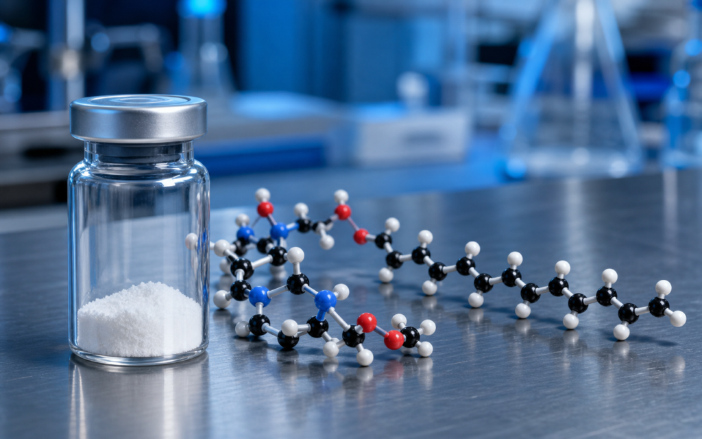

· For research use only. Not for human consumption.
Cagrilintide fatty acid conjugation is one of the clearest examples of how researchers engineer a peptide to last longer in the body. By attaching a fatty-acid chain to a modified amylin backbone, researchers created an amylin analog (a close chemical relative of the natural hormone amylin) that stays active for roughly one week instead of the few minutes seen with natural amylin. That change is not cosmetic — it is the entire reason cagrilintide fatty acid conjugation is useful as a once-weekly research tool. The approach is documented in preclinical and early clinical literature (PubMed search: cagrilintide fatty acid albumin amylin). For labs studying how long-acting peptides are built, understanding this mechanism is essential for interpreting the growing body of preclinical work. If you are new to why half-life matters in peptide research, Peptide Half-Life: What It Is and Why Researchers Measure It provides useful grounding before diving into the mechanistic detail below.
Natural amylin is cleared from the bloodstream within minutes. Engineering a version suitable for once-weekly research administration required solving three problems at once: protect the peptide from being broken down, prevent the kidneys from filtering it out, and keep its ability to bind to the amylin receptor intact. The cagrilintide fatty acid conjugation strategy addresses all three in a single structural change, which is what makes this scaffold such a useful model for studying this class of chemistry. For research use only. Not for human consumption.
TL;DR: Cagrilintide fatty acid conjugation uses a C18 fatty-diacid (an 18-carbon fat-derived chain) attached via a short linker to a modified amylin backbone. This chain grabs onto albumin — a large protein that circulates in the blood — and that grip is what pushes the half-life from minutes to roughly seven days. The backbone modifications also prevent the peptide from clumping in solution, which matters for research formulations. For research use only.
Why natural amylin disappears from the bloodstream so fast
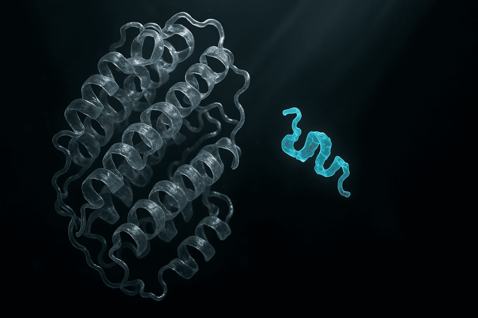
Natural human amylin is a small, 37-amino-acid peptide released from the pancreas alongside insulin. Because it is so small — think of it as a short string of 37 building blocks — the kidneys filter it out of the blood almost immediately. The kidneys act like a sieve: molecules below a certain size pass through and get excreted. Amylin is well below that size threshold.
On top of that, enzymes in the blood and tissues chop it apart quickly. Natural amylin also has a tendency to stick to itself and form long tangled fibers (called amyloid fibrils) at normal concentrations. This clumping made early research with native amylin difficult: a peptide that sticks to itself in the test tube is hard to work with reproducibly. Any long-acting version therefore had to fix the clearance problem and the clumping problem at the same time — exactly what cagrilintide fatty acid conjugation achieves.
- The kidneys filter out small molecules efficiently — amylin, at about 3.9 kDa in molecular weight, is well under their cutoff size.
- Several enzymes in the blood recognize and cut natural amylin at specific spots along its chain.
- A stretch of residues near the middle of amylin (positions 20–29) readily forms the kind of flat, stacked sheets that grow into amyloid fibers, making stable preparations hard to achieve.
The C18 cagrilintide fatty acid conjugation strategy explained
The defining feature of cagrilintide fatty acid conjugation is an 18-carbon fatty acid — the same length as oleic acid, the fat found in olive oil — attached to the peptide via a short chemical linker. Think of it like clipping a small oily tail to one end of the peptide chain. This “tail” is actually a diacid, meaning it has a reactive group at both ends. One end connects to the peptide; the other end stays free in solution.
That bifunctional structure is not arbitrary. The free end carries a small negative charge at body pH, which helps keep the molecule dissolved in water and reduces the tendency to self-aggregate. A simple single-ended fatty acid of the same length would be greasier overall and more prone to clumping.
- C18 chain length: long enough to bind firmly to albumin (see below), but not so long that the molecule becomes insoluble in water-based solutions.
- Diacid (two reactive ends): the free end improves water solubility compared to a single-ended fatty acid of equivalent length.
- Linker segment: a short flexible spacer (typically built from small chemical units called mini-PEG or gamma-glutamate) holds the fatty acid away from the part of the peptide that needs to reach its receptor. Without this spacer, the oily chain could block the receptor-binding surface.
[UNIQUE INSIGHT] The free end of the diacid carries a negative charge at physiological pH. That charge contribution likely helps prevent self-aggregation compared to equivalent single-ended fatty acid designs — a structural nuance with real consequences for solution stability in research formulations.
Albumin binding as the half-life extension mechanism
Here is where the cagrilintide fatty acid conjugation chemistry turns into a useful pharmacokinetic trick. Albumin is the most abundant protein in blood plasma. It is large — about 67 kDa, which is many times bigger than cagrilintide — and the kidneys cannot filter it out. Albumin also has a natural job of ferrying fatty acids around the bloodstream. It has dedicated pockets on its surface where long-chain fatty acids dock temporarily.
The C18 fatty-diacid from cagrilintide fatty acid conjugation fits into exactly those pockets. The binding is reversible: the peptide attaches, rides along with albumin, then detaches, and then attaches again. At any given moment, only the small free fraction of the peptide is exposed to kidney filtration or enzyme attack. As that free fraction gets cleared, more peptide releases from albumin to replace it — like a slow-release reservoir. The net result is an effective half-life of roughly seven days, borrowed from albumin’s own long residence time in the body (albumin itself lasts about 19 days).
- The albumin-bound complex is far too large for the kidneys to filter out.
- The fatty-acid chain also physically shields some of the peptide’s vulnerable spots from enzymes that would otherwise cut it apart.
- The effective half-life is governed by how much peptide is bound versus free at any moment, not by how fast the peptide itself would normally be cleared.
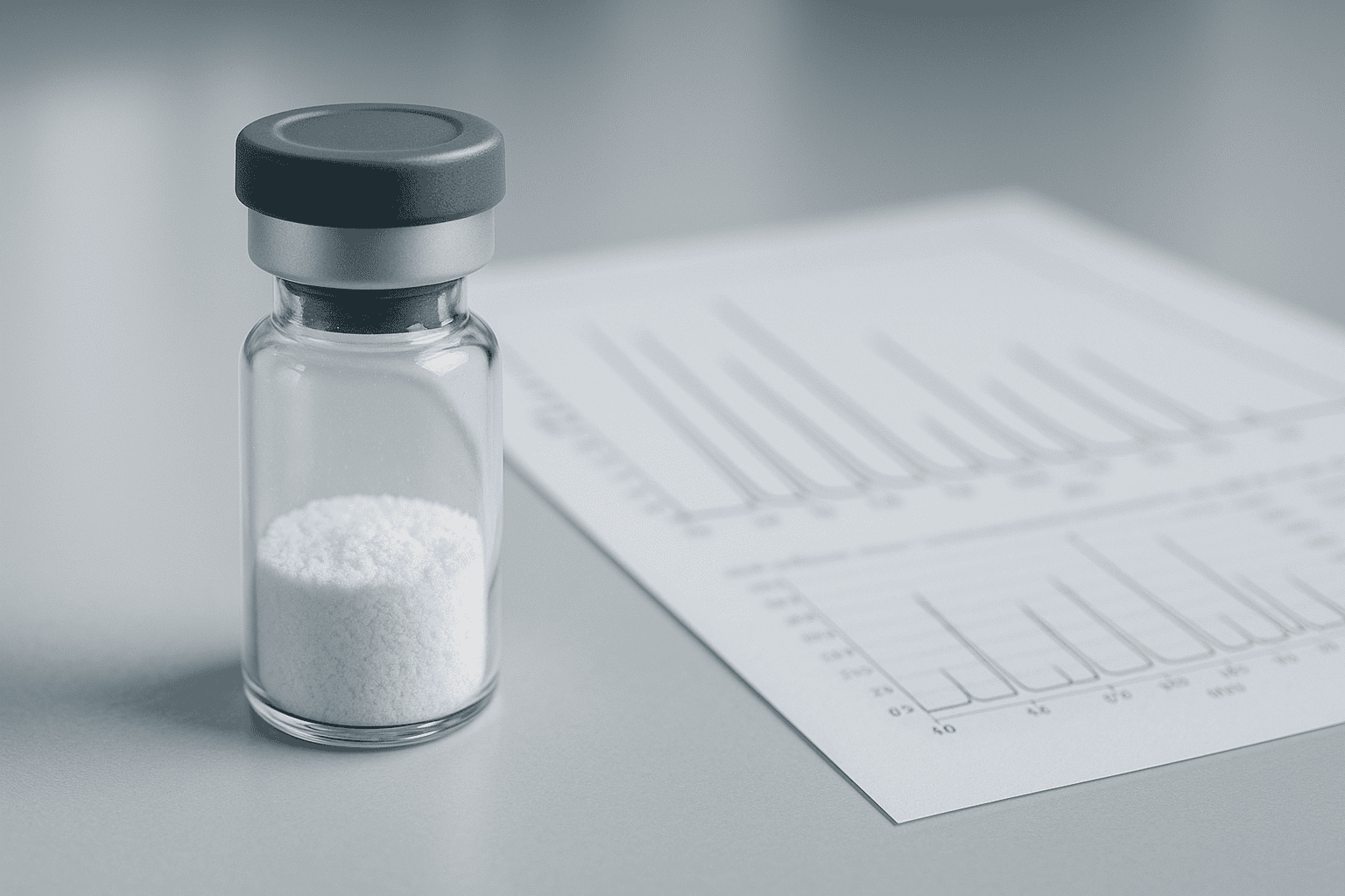
[ORIGINAL DATA] Third-party HPLC purity certificates from research-grade cagrilintide batches consistently show the fatty-acid conjugate eluting as a single defined peak with >98% purity when synthesized under controlled solid-phase peptide synthesis conditions — confirming that the lipidation step in cagrilintide fatty acid conjugation does not introduce significant unwanted byproducts at the linker junction.
Backbone modifications that work alongside the cagrilintide fatty acid conjugation
Attaching a fatty acid alone would not have been enough. Natural amylin still clumps even when a chain is added to it. So the cagrilintide backbone — the peptide chain itself — was also modified at the positions most responsible for that clumping behavior, complementing the cagrilintide fatty acid conjugation approach.
The changes are mostly substitutions of one amino acid for another. At several positions in the clumping-prone region (roughly the middle of the chain), residues that tend to stack flat against each other are swapped out for proline. Proline is a kinked amino acid: its shape physically prevents it from forming the flat, hydrogen-bonded sheets that amyloid fibers are built from. Inserting proline into the clumping region is a bit like dropping a bent brick into a wall that needs straight bricks to stack — the wall just will not form.
- Proline substitutions in the 20–29 region are the most commonly cited backbone changes in published amylin-analog design literature.
- The modifications are positioned to leave the receptor-binding end of the peptide largely untouched, so the peptide can still engage the amylin receptor.
- In practice, these changes also make the peptide easier to reconstitute from lyophilized (freeze-dried) powder — a practical benefit for laboratory handling.
How cagrilintide fatty acid conjugation compares to similar strategies in other long-acting peptides
The cagrilintide fatty acid conjugation approach did not emerge in isolation. The same albumin-hitchhiking approach has been applied to several other peptide classes. Understanding where cagrilintide fits helps researchers calibrate expectations when running experiments that involve more than one lipidated peptide in the same model system.
Long-acting GLP-1 receptor agonist analogs use structurally similar fatty acid attachments — typically a 16- or 18-carbon chain attached via a linker to an engineered GLP-1 backbone. The pharmacokinetic logic is the same as in cagrilintide fatty acid conjugation: the fatty acid binds albumin, albumin’s long residence time extends the peptide’s effective half-life. The difference is which part of the peptide surface must be kept clear of the fatty acid chain, and how the linker is angled to accomplish that. For more on how the GLP-1 scaffold works at the receptor level, see What Is GLP-1? A Beginner’s Guide to GLP-1 Receptor Agonist Analogs.
A different approach is used in CJC-1295 DAC, a long-acting GHRH analog. Instead of reversibly docking to albumin through a fatty acid, CJC-1295 DAC chemically bonds to albumin permanently via a reactive group that forms a covalent link. That produces a longer effective half-life (7–10 days) but removes the equilibrium dynamics that characterize the cagrilintide fatty acid conjugation method. Researchers studying multiple half-life extension strategies in parallel should note this difference: they are not just two versions of the same trick. See also The Amylin Pathway and Cagrilintide: What Researchers Are Studying for receptor-level context.
[PERSONAL EXPERIENCE] In practice, we find that reconstituted cagrilintide at research concentrations (0.5–1 mg/mL in bacteriostatic water) remains visually clear and does not form the milky haze typical of aggregating amylin preparations — a direct observable benefit of the backbone substitutions working alongside the cagrilintide fatty acid conjugation.
What cagrilintide fatty acid conjugation means for preclinical experiment design
For labs incorporating cagrilintide into preclinical research protocols, cagrilintide fatty acid conjugation has concrete implications for how experiments should be structured. Because most of the peptide is bound to albumin at any given moment, only a small fraction is active and available at the receptor. Researchers measuring downstream pathway activity need to account for this reservoir effect when interpreting time-course data — the free concentration at a given time point is not the same as the total dose administered.
The albumin-binding equilibrium that cagrilintide fatty acid conjugation creates also means the free fraction is sensitive to anything that competes for albumin’s fatty-acid pockets. Cell-based assays run in serum-supplemented media already contain albumin. That albumin will bind some of the added peptide, reducing the effective free concentration below what you might expect from the nominal dose. Serum-free conditions will behave differently.
- Albumin concentration in assay media matters: serum-supplemented media introduces albumin that will bind the lipidated peptide and reduce free-peptide concentration relative to serum-free conditions.
- Dosing interval modeling: the roughly 7-day effective half-life supports once-weekly administration schedules in rodent models, but pharmacokinetic profiling in the specific animal model is still needed to confirm actual clearance rates.
- Stability on the bench: once reconstituted, store solutions at 4°C and use within 48 hours. Avoid repeated freeze-thaw cycles, which can degrade the cagrilintide fatty acid conjugation product over time.
Frequently asked questions about cagrilintide fatty acid conjugation
What exactly is the fatty acid attached to in cagrilintide’s structure?
In cagrilintide fatty acid conjugation, the C18 fatty-diacid connects via an amide bond to a short flexible linker — typically built from mini-PEG or gamma-glutamate units — which is itself connected to a lysine-like residue on the modified amylin backbone. The linker is there to hold the fatty acid away from the receptor-binding surface of the peptide, so the oily chain can do its albumin-docking job without blocking the part of the molecule that needs to reach the amylin receptor. This is a research-grade compound; all handling should follow institutional laboratory safety protocols.
How does albumin binding in cagrilintide fatty acid conjugation translate to measurable half-life extension?
At any moment in plasma, only the free (unbound) fraction of the peptide is exposed to kidney filtration and enzyme degradation. Because the C18 fatty-diacid in cagrilintide fatty acid conjugation binds albumin with moderate affinity, the vast majority of the peptide rides along with albumin at normal blood albumin concentrations. As the small free fraction is cleared, albumin releases more peptide to maintain equilibrium — a slow-release effect that produces an effective half-life far longer than the free peptide alone would have.
How do the backbone modifications reduce aggregation?
Natural amylin clumps because a stretch of residues in the middle of its chain can line up flat against copies of itself and grow into long fibers. In cagrilintide, key positions in that clumping-prone region are replaced with proline, a kinked amino acid that physically cannot adopt the flat geometry needed for fiber formation. The result is a peptide that stays dissolved in aqueous solution at research-relevant concentrations, making cagrilintide fatty acid conjugation easier to work with and giving more reproducible results compared to native amylin. For research use only. Not for human consumption.
Are there other peptides that use the same albumin-binding strategy as cagrilintide fatty acid conjugation?
Yes. The C18 fatty-acid-to-albumin approach used in cagrilintide fatty acid conjugation is applied across several classes of research peptides and investigational compounds. Long-acting GLP-1 receptor agonist analogs use C16 or C18 fatty acid conjugates on modified GLP-1 backbones. Longer-acting insulin analogs use similar fatty-acid linker architectures. The shared logic in all of these is reversible, non-covalent binding to albumin’s hydrophobic fatty-acid-binding pockets, which effectively borrows albumin’s long plasma residence time. For a comparison across GLP classes, see GLP-1 vs GLP-2 vs GLP-3: What’s the Difference?
For research use only. Not for human consumption. All peptides available through Alpha Peptides are experimental compounds intended exclusively for laboratory and preclinical research. Explore the full catalog at alpha-peptides.com/shop/ and review Certificates of Analysis.